Neuron Draw And Label
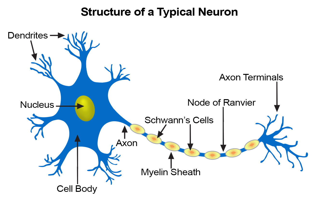
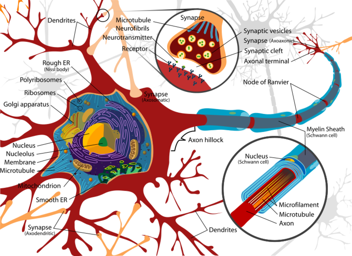
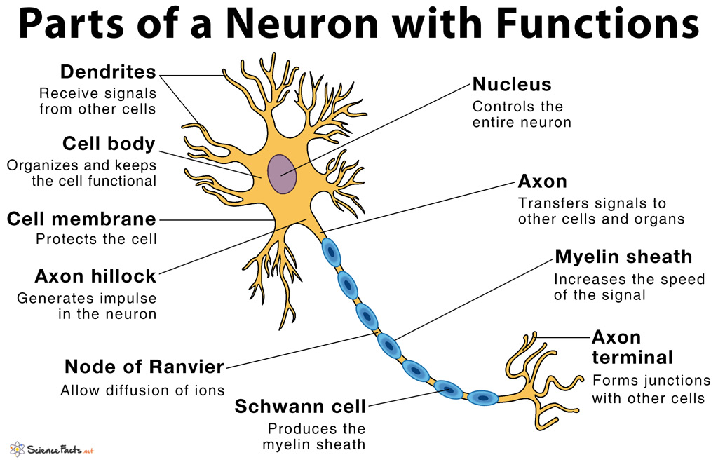
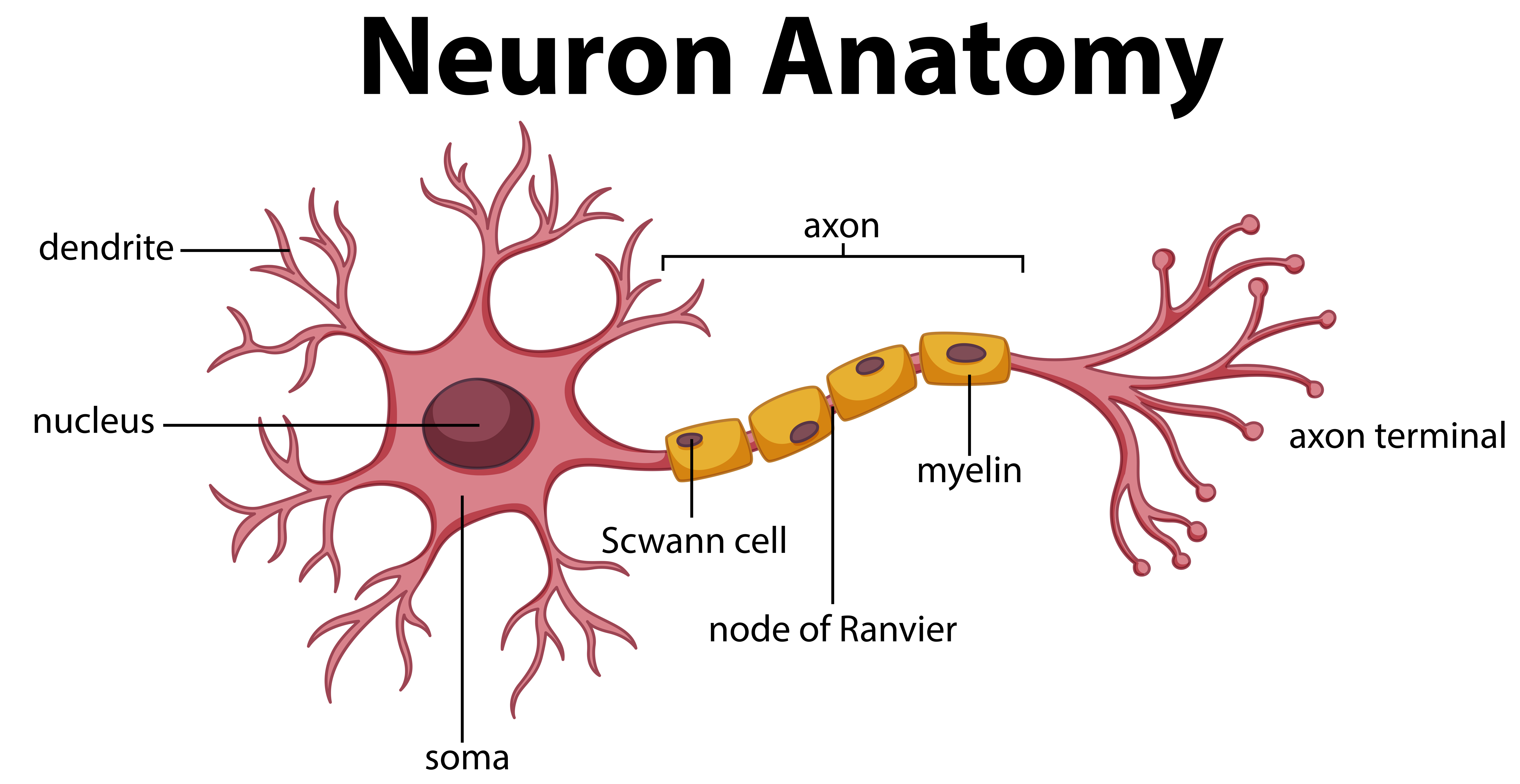
Neuron Draw And Label - (8) this neuron part gives messages to muscle tissue. Web draw and label a myelinated neuron showing the cell body, dendrite, axon, axon terminal, schwann cells and nodes of ranvier. A neuron is made up of three regions, namely the nerve cell body (soma), axon and dendrite. This is a tapering of the cell body toward the axon fiber. These cells pass signals from the outside of your body, such as touch, along to the central nervous system.
Choose the correct names for the parts of the neuron. These cells pass signals from the outside of your body, such as touch, along to the central nervous system. Web diagram of neuron with labels here is the description of human neuron along with the diagram of the neuron and their parts. The axon and dendrite are the filamentous structures of the neuron. Web neurons communicate with one another at junctions called synapses. In these synapses, ions flow directly between cells. The anatomy of a neuron neurons are a significant part of the nervous system.
Describe the Anatomy of a Neuron
Read the definitions, then label the neuron diagram below. And it will teach you to draw the neuron very easily. Web diagram of neuron with labels here is the description of human neuron along with.
Anatomy Of A Neuron ANATOMY
Read the definitions, then label the neuron diagram below. At a synapse, one neuron sends a message to a target neuron—another cell. Web neuroglia labeling with google slides save paper by assigning labeling worksheets on.
What Is a Neuron? Diagrams, Types, Function, and More
Dendrite = receives chemical signals from other Bipolar neurons have one axon and only one dendrite branch. Read the definitions, then label the neuron diagram below. Start studying label parts of a neuron. Web neurons.
FileNeuron1.jpg Simple English Wikipedia, the free encyclopedia
And it will teach you to draw the neuron very easily. Web this article introduces how to understand neurons and the functions with neurons labeled diagrams. All neurons have three main parts. (8) this neuron.
Draw A Labelled Diagram Showing The Structure Of A Neuron Biology
Web carries the cell's impulse to the terminal. These cells pass signals from the outside of your body, such as touch, along to the central nervous system. Dendrite = receives chemical signals from other Where.
Structure of a Neuron Owlcation
Neurons, also known as nerve cells, are essentially the cells that make up the brain and the nervous system. Provide a brief description of the function of each labeled structure beside its label. A neuron.
Neuron Study Guide Inspirit Learning Inc
Bipolar neurons have one axon and only one dendrite branch. Spreads the cell's impulse out to reach other neurons. Web unipolar neurons are also known as sensory neurons. Send electrical impulses to neighboring neurons. It.
neuron diagram labeled
The anatomy of a neuron neurons are a significant part of the nervous system. A neuron is made up of three regions, namely the nerve cell body (soma), axon and dendrite. (8) this neuron part.
How TO Draw neuron step by step easy/diagram of neuron/neuron drawing
Web the parts of the neuron have been labeled. Where the axon emerges from the cell body, there is a special region referred to as the axon hillock. And it will teach you to draw.
Neuron Diagram Straight from a Scientist
Web anatomy of a neuron google classroom about transcript neurons (or nerve cells) are specialized cells that transmit and receive electrical signals in the body. Cell body each neuron has a cell body with a.
Neuron Draw And Label The anatomy of a neuron neurons are a significant part of the nervous system. (1) (2) (3) (4) (5) (6) this neuron part receives messages from other neurons. Start studying label parts of a neuron. If not, then it’s time to learn! Web parts of a neuron diagram.


