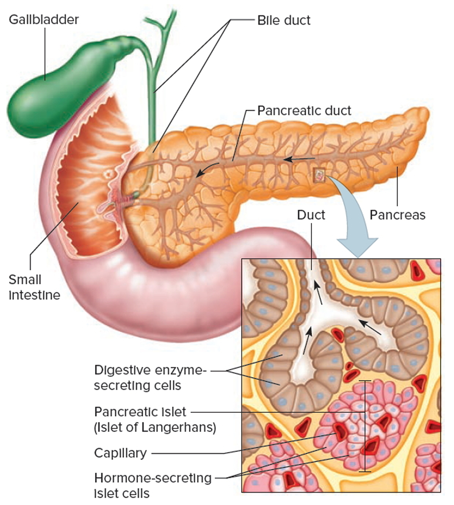
Pancreas Drawing
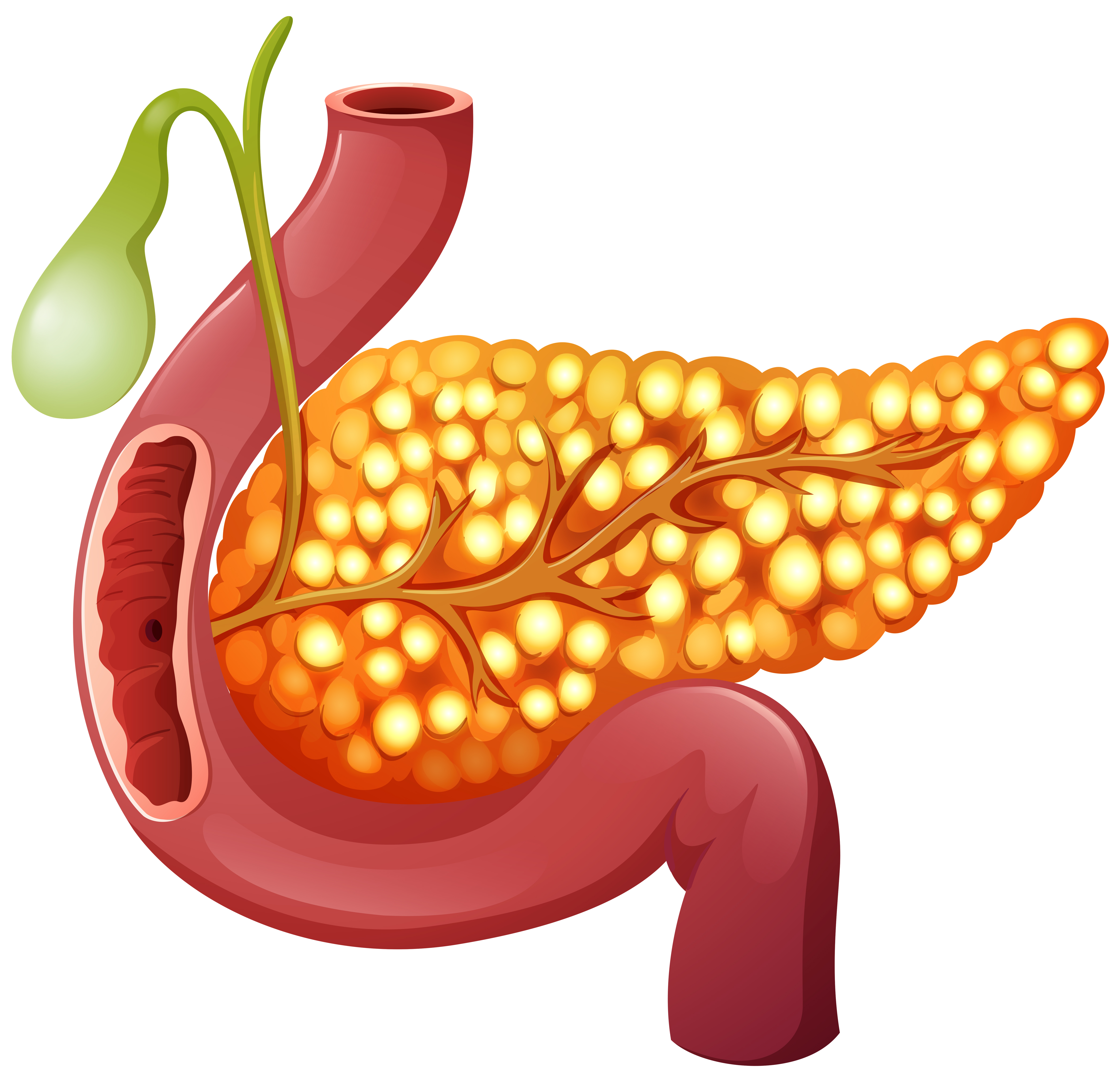
Pancreas Drawing - Conditions that affect the pancreas range from type 1 diabetes and type 2 diabetes to pancreatitis and pancreatic cancer. Secreting enzymes that aid in digestion and releasing hormones, in particular insulin, to help regulate the amount of glucose (sugar) in the blood). It is around 12 to 15 cm long and 4 cm wide, and sits across the lumbar spine. Serous acini with zymogenic cells of exocrine part #4. Drawing shows the pancreas, stomach, spleen, liver, bile ducts, gallbladder, small intestine, and colon.
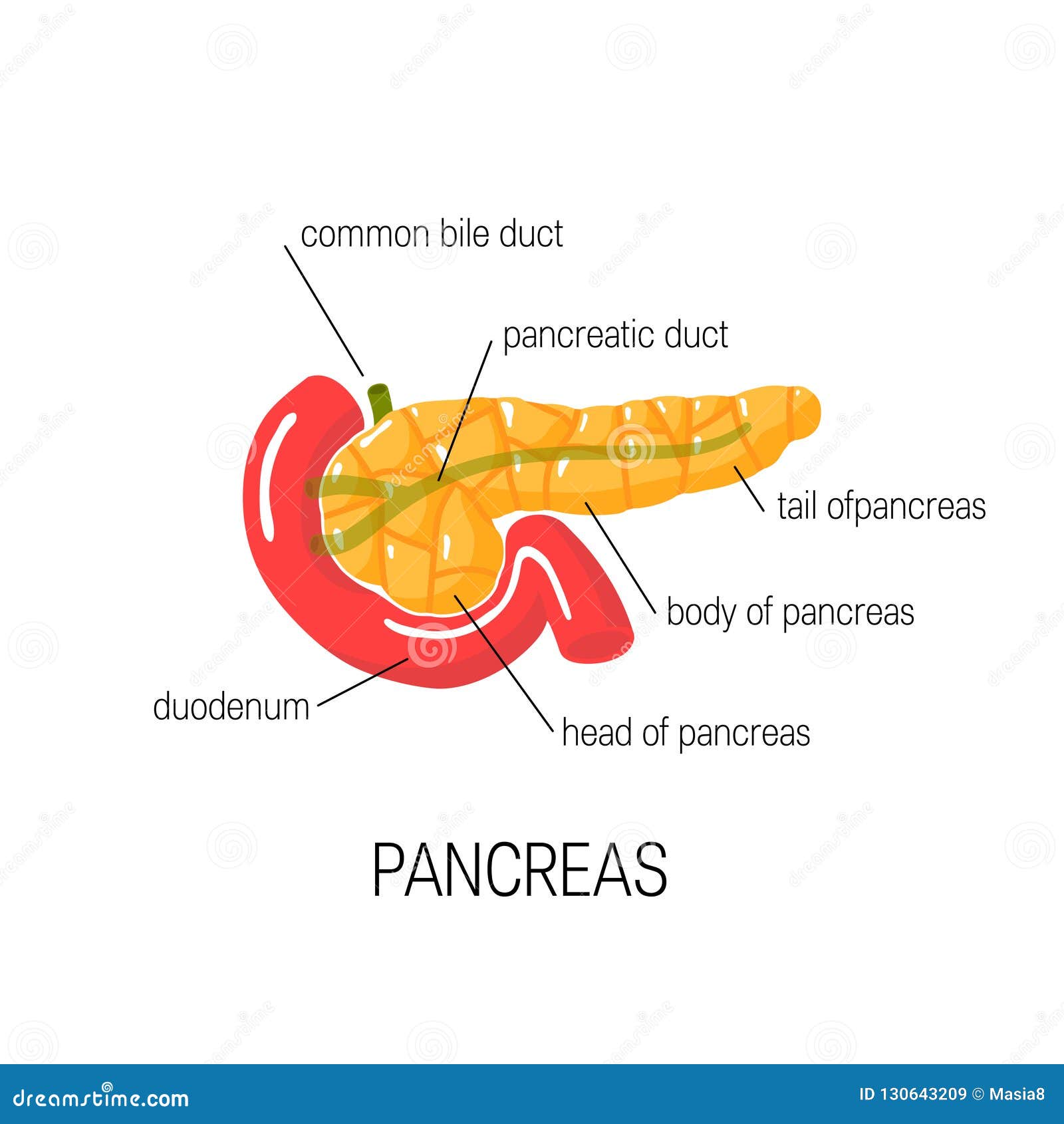
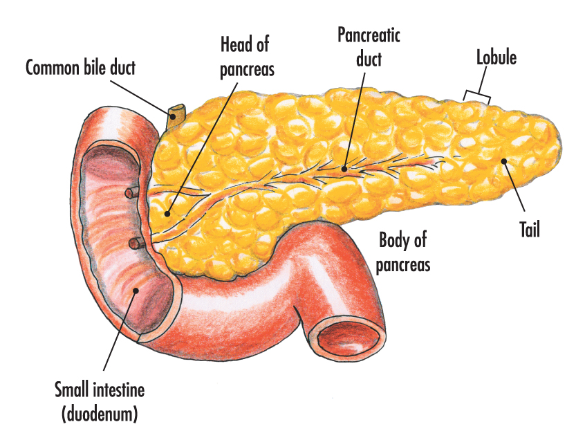
It produces substances that impact digestion and blood sugar. Figures 1a and 1b are two normal human pancreases from autopsies of adults. This cartoon represents the anatomical features of a “slice” of the abdomen at the level depicted in the upper right hand corner of the figure. The head, the body, and the tail. Chapter 1 of the fascicle is recommended as a source for additional detail regarding pancreatic anatomy and histology, and for discussion of the genetic control of pancreatic development. Interlobular connective tissue septa of pancreas #3. The bile duct and pancreatic duct are also shown.

Human Pancreas drawing How to draw human Pancreas Human pancreas drawing step by stepEasy
Web how to draw pancreas | step by step drawing adimu show 43.4k subscribers join subscribe subscribed 130 share save 13k views 1 year ago #pancreas #howtodraw #adimushow #pancreas #howtodraw. You can take steps to.
Pancreas Location, Anatomy and Function in Digestion
Anterior to the pancreas are the stomach, colon, omentum. The two photos illustrate that there is considerable individual variation in the shape of the pancreas. Conditions that affect the pancreas range from type 1 diabetes.
A healthy human Pancreas 303570 Vector Art at Vecteezy
The gross anatomy of the human pancreas can vary. 99% of the pancreas is exocrine and 1% is endocrine. The pancreas is both an exocrine accessory digestive organ and a hormone secreting endocrine gland. Anterior.

Pin on Diagrams and Infographics
The head, the body, and the tail. Web anatomy of the pancreas; Drawing shows the pancreas, stomach, spleen, liver, bile ducts, gallbladder, small intestine, and colon. Web the pancreas is an organ of the digestive.
Medical Diagram of Pancreas, Vector Illustration Stock Vector Illustration of endocrine
It has two important functions: You can take steps to help keep your pancreas healthy, including maintaining a healthy diet and weight. The pancreas has three parts: The bulk of the pancreatic tissue is formed.

Illustration of the normal pancreatic anatomy. Pancreas is divided into... Download Scientific
Conditions that affect the pancreas range from type 1 diabetes and type 2 diabetes to pancreatitis and pancreatic cancer. The images range from classic work of skilled medical artists to original drawings and photomicrographs from.

Illustration of a woman's pancreas Stock Image F023/5801 Science Photo Library
It is customary to refer to various portions of the pancreas as head, body, and tail. Liver insulin diabetes pancreatic cancer varicose veins pancreas icon pancreas illustration pancreas cancer The two photos illustrate that there.
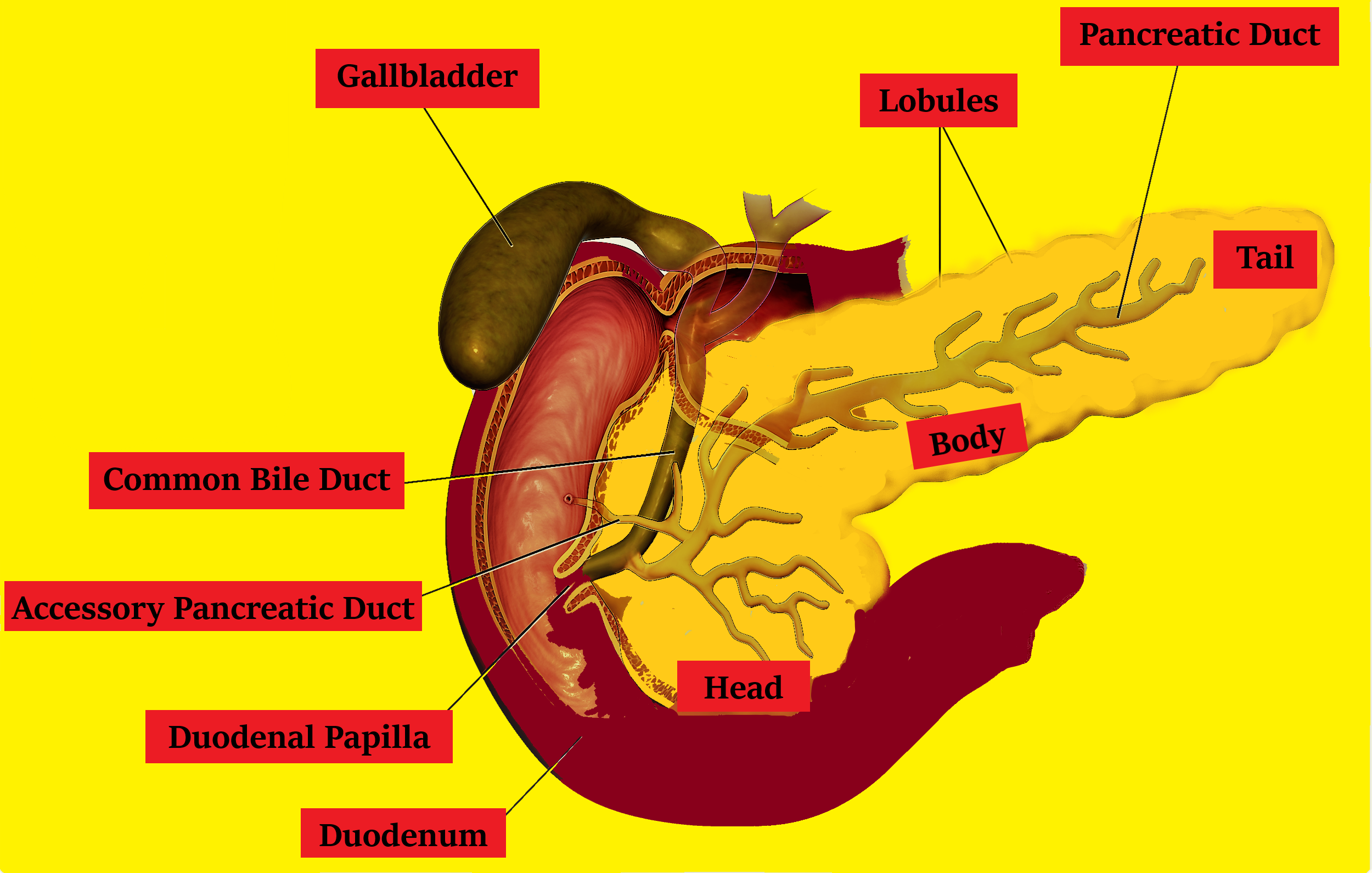
Draw a neat labeled diagram of the pancreas with their associated structure.
The bulk of the pancreatic tissue is formed by the exocrine component, which consists of many serous pancreatic acini cells. It has two important functions: Secreting enzymes that aid in digestion and releasing hormones, in.

Contour vector outline drawing of human pancreas organ. Medical design editable template Stock
Interlobular connective tissue septa of pancreas #3. Anterior to the pancreas are the stomach, colon, omentum. It has two important functions: The bile duct and pancreatic duct are also shown. Web pancreas anatomy and human.
The pancreas Anatomy of the pancreas Structure of the pancreas
The two photos illustrate that there is considerable individual variation in the shape of the pancreas. Pancreas in situ seen from the anterior view. Drawing shows the pancreas, stomach, spleen, liver, bile ducts, gallbladder, small.
Pancreas Drawing Web anatomical structure the pancreas is typically divided into five parts: Chapter 1 of the fascicle is recommended as a source for additional detail regarding pancreatic anatomy and histology, and for discussion of the genetic control of pancreatic development. The images range from classic work of skilled medical artists to original drawings and photomicrographs from leaders in the study of pancreatic anatomy. Interlobular connective tissue septa of pancreas #3. Web anatomy function associated conditions tests the pancreas is a gland located deep inside the abdomen, just behind the lower part of the stomach.