10 Heart Perfusion Study Tips For Better Diagnosis

The field of cardiology has witnessed significant advancements in diagnostic techniques, and one such crucial method is heart perfusion study. This non-invasive test is pivotal in assessing the blood flow to the heart muscle, thereby helping in the diagnosis of coronary artery disease (CAD) and other heart conditions. To ensure accurate and reliable results from a heart perfusion study, healthcare professionals and patients alike must be aware of certain key aspects. Here are 10 essential tips for a better diagnosis:
Preparation is Key: Before undergoing a heart perfusion study, patients should be well-prepared. This includes avoiding heavy meals, especially those high in fat, for at least 4 hours prior to the test. Caffeine and nicotine should also be avoided for a few hours before the test, as they can interfere with the heart rate and blood pressure, potentially affecting the test results.
Understand the Procedure: It’s crucial for patients to have a clear understanding of what the procedure entails. A heart perfusion study, also known as a myocardial perfusion scan, involves the use of a small amount of radioactive material (tracer) that is injected into a vein in the arm. This tracer travels to the heart and releases signals that are picked up by a special camera, creating images of the heart muscle.
Choose the Right Tracer: The choice of tracer can significantly affect the quality of the images obtained. Technetium-99m sestamibi (MIBI) and Thallium-201 are common tracers used. Each has its own set of advantages and disadvantages, and the choice between them often depends on the patient’s specific condition and the availability of the tracers.
Exercise Stress Test: For many patients, a heart perfusion study is conducted in conjunction with an exercise stress test. This involves walking on a treadmill or pedaling a stationary bike to increase the heart rate and simulate the effects of exercise on the heart. The ability to perform physical exercise to an adequate level is crucial for the success of this part of the test.
Pharmacological Stress Test Alternative: For patients who cannot perform an exercise stress test due to physical limitations, a pharmacological stress test can be used as an alternative. This involves the use of medications like adenosine or regadenoson to dilate the blood vessels and increase blood flow to the heart, simulating the effects of exercise.
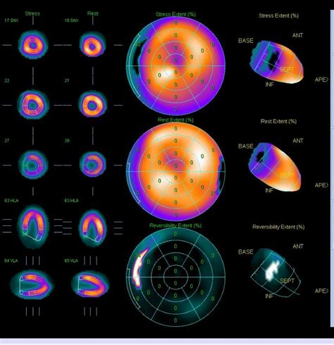
Image Interpretation: The interpretation of the images obtained from a heart perfusion study requires a high level of expertise. Healthcare professionals should look for areas of the heart muscle that do not receive adequate blood flow, which could indicate blockages in the coronary arteries. The use of advanced software and techniques, such as gated SPECT, can enhance the accuracy of image interpretation.
Combination with Other Diagnostic Tools: For a more comprehensive diagnosis, a heart perfusion study can be combined with other diagnostic tools like echocardiography, cardiac catheterization, or cardiac MRI. Each of these tests provides different information, and together they can offer a complete picture of the heart’s condition.
Patient Cooperation: The success of a heart perfusion study heavily relies on patient cooperation. Patients must be able to follow instructions accurately, such as holding still during the scanning process and breathing normally. Any movement can blur the images, making them less useful for diagnostic purposes.
Addressing Anxiety and Fear: Undergoing any medical test can be a source of anxiety for patients. It’s essential for healthcare providers to address these concerns, explaining the procedure in detail, discussing potential risks and benefits, and ensuring that the patient feels as comfortable as possible throughout the process.
Follow-Up and Lifestyle Changes: After the diagnosis, it’s crucial for patients to understand the implications of the results and the necessary follow-up actions. Depending on the findings, this might include lifestyle changes such as adopting a healthier diet, increasing physical activity, quitting smoking, and managing stress. In cases where coronary artery disease is diagnosed, further treatment such as medication, angioplasty, or bypass surgery might be recommended.
In conclusion, a heart perfusion study is a valuable diagnostic tool that, when used correctly and in conjunction with other tests, can provide critical information about the heart’s condition. By understanding and implementing these tips, healthcare professionals can ensure that their patients receive the most accurate and beneficial results from this test, ultimately leading to better diagnosis and treatment outcomes.
FAQ Section
What is the primary purpose of a heart perfusion study?
+The primary purpose of a heart perfusion study is to assess the blood flow to the heart muscle, helping in the diagnosis of coronary artery disease (CAD) and other heart conditions by identifying areas of the heart that do not receive adequate blood flow.
How long does a heart perfusion study typically take?
+A heart perfusion study can take approximately 2 to 3 hours to complete, including preparation time, the actual scanning, and image interpretation. However, the duration may vary depending on the specific procedures involved and the patient’s condition.
Are there any risks associated with a heart perfusion study?
+While generally considered safe, a heart perfusion study involves exposure to small amounts of radiation from the tracer. The risks associated with this radiation exposure are minimal, but it’s essential to discuss any concerns with a healthcare provider, especially for pregnant women or those with certain medical conditions.
Can a heart perfusion study detect other heart conditions besides CAD?
+Yes, besides CAD, a heart perfusion study can provide insights into other heart conditions such as cardiomyopathy, where the heart muscle becomes thickened or stiff, affecting its ability to pump blood efficiently. It can also be used to assess the viability of heart muscle after a heart attack.
How often can a heart perfusion study be repeated?
+The frequency of repeating a heart perfusion study depends on the patient’s condition and the reason for the follow-up. Generally, to minimize radiation exposure, healthcare providers try to limit the number of tests. However, if there are significant changes in symptoms or if the patient undergoes interventions like angioplasty or bypass surgery, a follow-up study may be necessary to assess the outcomes of these treatments.
Is a heart perfusion study covered by insurance?
+Coverage for a heart perfusion study varies by insurance provider and the patient’s specific policy. Generally, if the test is deemed medically necessary by a healthcare provider, it is likely to be covered. However, patients should check with their insurance company beforehand to understand their coverage and any out-of-pocket costs they might incur.


