12 Cat Scan Facts That Improve Diagnosis

The evolution of medical imaging has been a pivotal factor in the advancement of diagnostic capabilities, and one of the most influential technologies in this realm is the Cat Scan, more formally known as Computed Tomography (CT). This sophisticated imaging modality has revolutionized the field of medicine by providing high-resolution cross-sectional images of the body, thereby improving the accuracy and speed of diagnoses. Here are 12 critical facts about Cat Scans that highlight their role in enhancing diagnostic outcomes:
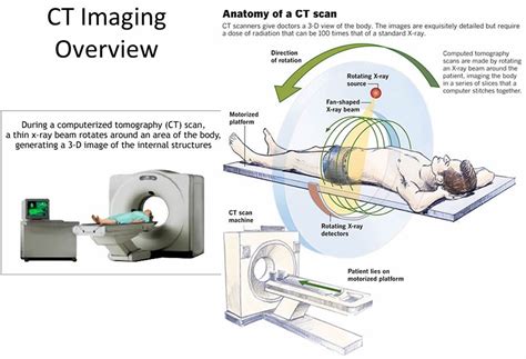
Basic Principle: A Cat Scan works on the principle of tomography, which involves the use of computer-processed combinations of multiple X-ray measurements taken from different angles to produce cross-sectional images of the body. These images allow for the visualization of internal structures in great detail, facilitating the detection of abnormalities that might not be visible through conventional X-rays.
History and Development: The invention of the CT scan is credited to Sir Godfrey Hounsfield and Allan McLeod Cormack, who were awarded the Nobel Prize in Physiology or Medicine in 1979 for their pioneering work. Since its introduction in the early 1970s, CT technology has undergone significant advancements, leading to improved image quality, faster scanning times, and reduced radiation doses.
Applications in Diagnosis: CT scans are versatile diagnostic tools used in various medical specialties, including neurology, cardiology, oncology, and emergency medicine. They are particularly useful for diagnosing conditions such as strokes, cancers, vascular diseases, and injuries, especially those involving internal organs and structures.
Contrast Agents: To enhance the visibility of certain tissues or structures, contrast agents may be administered before or during the scan. These substances can be given orally or intravenously and work by absorbing X-rays, thereby increasing the contrast between different types of tissues in the images produced. This can be particularly helpful in highlighting tumors, blood vessels, or areas of inflammation.
Radiation Exposure: One of the concerns associated with CT scans is the exposure to ionizing radiation, which carries a small risk of inducing cancer. However, technological advancements have led to the development of low-dose protocols that minimize radiation exposure while maintaining image quality. Patients should discuss the benefits and risks of CT scans with their healthcare providers, especially if they require multiple scans over time.
Speed and Convenience: Modern CT scanners are capable of imaging the entire body in a matter of seconds, making them invaluable in emergency situations where time is critical. The speed and convenience of CT scans also reduce the need for sedation, especially in pediatric or claustrophobic patients, as the scanning process is quick and relatively comfortable.
High-Resolution Imaging: The resolution of CT scans has improved significantly over the years, allowing for the detection of smaller lesions and more precise delineation of anatomical structures. This high-resolution imaging capability is crucial for planning treatments, such as surgery or radiation therapy, and for monitoring disease progression or response to treatment.
3D Reconstruction: One of the advanced features of contemporary CT scanners is their ability to reconstruct images in three dimensions. This capability provides a more comprehensive understanding of complex anatomical relationships and is particularly useful in preoperative planning, allowing surgeons to visualize the surgical site in detail before making an incision.
Dual-Energy CT: Dual-energy CT is an advanced scanning technique that uses two different X-ray energies to produce images. This technology enhances material differentiation, allowing for better detection of certain types of lesions, such as those containing iodine or calcium, and can reduce the need for additional imaging tests.
Cardiac CT: Cardiac CT scans are specialized for imaging the heart and its vessels. They are used for detecting coronary artery disease, assessing cardiac function, and evaluating the heart’s structure. The high-speed nature of modern CT scanners makes them ideal for cardiac imaging, as they can capture images of the heart between beats, reducing motion artifact.
CT Angiography: CT angiography involves the use of contrast agents to visualize the blood vessels and diagnose vascular conditions such as aneurysms, stenosis, or malformations. This non-invasive procedure offers a safer alternative to traditional angiography and can provide critical information for treatment planning.
Future Developments: The future of CT scan technology holds promise for even further improvements in image quality, reduction in radiation exposure, and expansion of applications. Advances in artificial intelligence and machine learning are expected to play a significant role in enhancing image analysis, potentially leading to automated detection of abnormalities and personalized medicine approaches.
In conclusion, the Cat Scan has revolutionized the field of diagnostic imaging, offering healthcare providers a powerful tool for visualizing internal body structures and diagnosing a wide range of medical conditions. As technology continues to evolve, we can expect to see even more innovative applications of CT scans in the pursuit of improved patient outcomes and personalized care.
What is the primary difference between a CT scan and a traditional X-ray?
+The primary difference lies in the ability of a CT scan to produce cross-sectional images of the body, allowing for the detailed visualization of internal structures, whereas traditional X-rays provide two-dimensional images.
How does a CT scan help in the diagnosis of cancers?
+CT scans play a crucial role in cancer diagnosis by providing detailed images that help identify tumors, their size, location, and whether they have spread to other parts of the body. This information is vital for staging the cancer and planning appropriate treatment.
What safety precautions are taken to minimize radiation exposure during a CT scan?
+To minimize radiation exposure, CT scans are designed to use the lowest possible dose necessary to produce high-quality images. Additionally, protocols such as low-dose scanning for children and the use of iterative reconstruction techniques to reduce noise and allow for lower doses are implemented.
Can CT scans be used for patients with claustrophobia or other anxieties related to enclosed spaces?
+Yes, CT scans can be adapted for patients with claustrophobia or other anxieties. Open-bore CT scanners, which have a larger opening, can be less intimidating. Additionally, Healthcare providers may offer sedation or use other relaxation techniques to make the scanning process more comfortable for anxious patients.
How often should a patient undergo a CT scan for monitoring a condition?
+The frequency of undergoing a CT scan for monitoring a condition depends on the specific medical condition, its severity, and the treatment plan. Healthcare providers determine the appropriate scanning schedule based on individual patient needs, balancing the benefits of regular monitoring against the risks associated with radiation exposure.
Are CT scans covered by health insurance?
+Generally, CT scans that are deemed medically necessary are covered by health insurance plans. However, coverage can vary depending on the insurance provider, the specific policy, and the reason for the scan. It's essential for patients to verify their coverage before undergoing a CT scan.
The information provided here underscores the significant role that CT scans play in modern medicine, offering a powerful diagnostic tool that can lead to more accurate diagnoses and, ultimately, better patient outcomes. By understanding the capabilities, limitations, and appropriate uses of CT scans, healthcare providers and patients alike can make informed decisions about when and how to utilize this technology.


