Anatomy Scan 20 Weeks

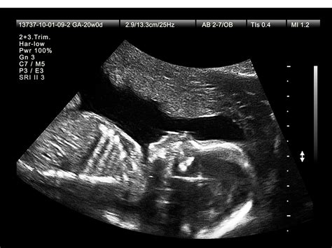
The 20-week anatomy scan, also known as the anatomy ultrasound or level II ultrasound, is a pivotal moment in pregnancy. This comprehensive scan is typically performed between 18 and 22 weeks of gestation, with 20 weeks being the most common timeframe. The primary goal of this scan is to assess the fetal anatomy in detail, evaluating the development and structure of various organs and body systems. This information is crucial for identifying any potential anomalies or issues that may require closer monitoring or intervention.
Preparation for the Scan
Before undergoing the 20-week anatomy scan, it’s essential for expectant mothers to understand the procedure and how to prepare. This includes:
- Hydration: Drinking plenty of water before the scan can help improve the quality of the ultrasound images by filling the bladder, which acts as a window for better visualization of the fetus.
- Full Bladder: A full bladder is often required for transabdominal ultrasound scans. However, the specific requirements may vary depending on the clinic or preferences of the sonographer.
- Comfortable Clothing: Wearing loose, comfortable clothing can make the scanning process easier and more comfortable.
The Scanning Process
The 20-week anatomy scan is performed by a trained sonographer who uses a transabdominal ultrasound machine. The process involves:
- Application of Gel: A clear gel is applied to the abdomen to help the ultrasound waves pass through more efficiently.
- Probe Movement: The sonographer moves a probe (transducer) over the abdomen, capturing images of the fetus from various angles.
- Image Analysis: The sonographer analyzes the images in real-time, taking measurements and assessing the development of the fetus.
What to Expect During the Scan
During the 20-week anatomy scan, the sonographer will examine the fetus’s major organs and body structures, including:
- Brain and Skull: Evaluating for any abnormalities in brain development or skull structure.
- Face and Neck: Checking for cleft lip or palate and ensuring proper development of the eyes, nose, and mouth.
- Heart: Assessing the heart’s structure and function, including the chambers and major blood vessels.
- Abdomen: Examining the liver, stomach, kidneys, and intestines.
- Skeletal System: Evaluating the development of the arms, legs, hands, and feet.
- Genitalia: Determining the sex of the baby, if desired, and assessing for any abnormalities.
After the Scan
Following the 20-week anatomy scan, the sonographer or a radiologist will review the images and measurements taken during the scan. This information is then discussed with the expectant mother, highlighting any findings that may be concerning. If any issues are detected, further testing or monitoring may be recommended.
FAQs
What is the purpose of a 20-week anatomy scan?
+The primary purpose is to evaluate the fetus's anatomy, checking for any developmental issues or abnormalities that may require medical attention.
Can the sex of the baby be determined during the scan?
+Yes, in most cases, the sex of the baby can be determined during the 20-week anatomy scan, provided the baby is in a favorable position.
What happens if an issue is found during the scan?
+If an issue is detected, the expectant mother may be referred for further testing, such as an amniocentesis or a more detailed fetal echo, and will be counseled on the findings and any necessary next steps.
Conclusion
The 20-week anatomy scan is a critical milestone in pregnancy, offering a detailed assessment of the fetus’s development and anatomy. While it can provide valuable insights and reassurance, it’s also a moment when potential issues may be identified, allowing for timely intervention. Understanding the purpose, process, and potential outcomes of this scan can help expectant mothers navigate this period with greater knowledge and peace of mind.


