Computed Tomography Brain Scan

The human brain, a complex and intricate organ, has long been a subject of fascination for medical professionals and researchers alike. One of the most significant advancements in understanding brain function and structure has been the development of computed tomography (CT) brain scans. This non-invasive imaging technique has revolutionized the field of neurology, enabling doctors to diagnose and treat a wide range of brain-related conditions with unprecedented accuracy.
History of CT Brain Scans
The first CT scan was performed in 1971 by Godfrey Hounsfield and Allan McLeod Cormack, who were later awarded the Nobel Prize in Physiology or Medicine for their groundbreaking work. The initial CT scanners used a combination of x-rays and computer algorithms to produce cross-sectional images of the brain. Over the years, CT technology has undergone significant improvements, with the introduction of faster scanning times, higher resolution images, and advanced reconstruction algorithms.
How CT Brain Scans Work
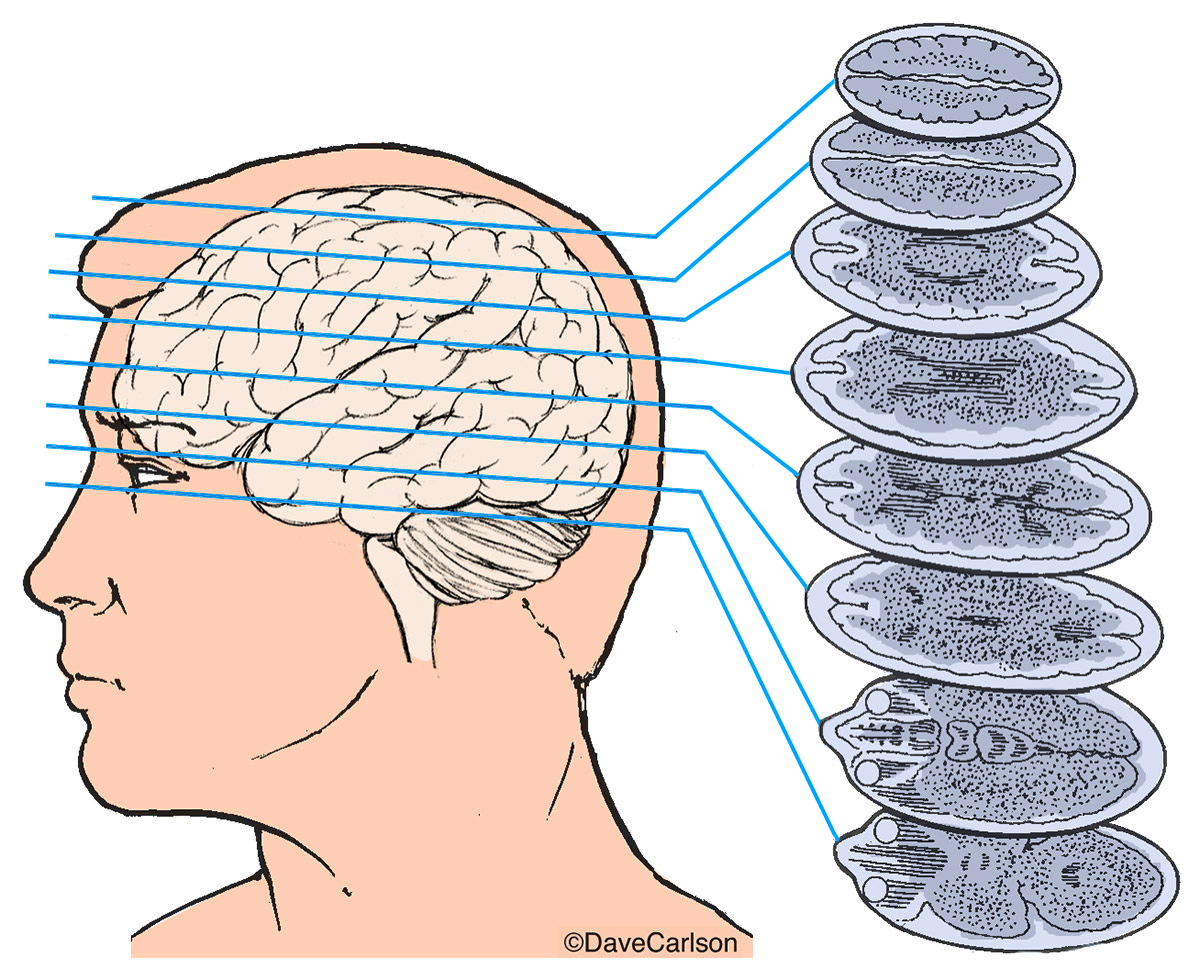
A CT brain scan typically begins with the patient lying on a movable table that slides into a large, doughnut-shaped machine. The scanner uses a series of x-ray beams to capture images of the brain from different angles. The x-rays pass through the brain, and the resulting images are reconstructed into a three-dimensional picture using sophisticated computer software. The entire process usually takes between 10-30 minutes, depending on the type of scan and the patient’s condition.
Types of CT Brain Scans
There are several types of CT brain scans, each designed to provide specific information about brain structure and function. Some of the most common types include:
- Non-contrast CT scan: This is the most basic type of CT scan, which uses x-rays to produce images of the brain without the use of intravenous contrast agents.
- Contrast-enhanced CT scan: This type of scan uses a contrast agent, usually iodine or barium, to highlight specific areas of the brain, such as blood vessels or tumors.
- CT angiography: This scan uses a contrast agent to visualize the blood vessels in the brain, helping doctors diagnose conditions such as aneurysms or arteriovenous malformations.
- CT perfusion scan: This scan measures blood flow to the brain, helping doctors diagnose conditions such as stroke or cerebral vasculitis.
Advantages of CT Brain Scans
CT brain scans offer several advantages over other imaging modalities, including:
- Speed: CT scans are generally faster than other imaging modalities, such as magnetic resonance imaging (MRI) or positron emission tomography (PET) scans.
- Availability: CT scanners are widely available in hospitals and imaging centers, making them a convenient option for patients.
- Cost-effectiveness: CT scans are often less expensive than other imaging modalities, making them a cost-effective option for patients and healthcare providers.
- High-resolution images: CT scans produce high-resolution images of the brain, allowing doctors to diagnose conditions with unprecedented accuracy.
Clinical Applications of CT Brain Scans
CT brain scans have a wide range of clinical applications, including:
- Diagnosing stroke: CT scans can quickly diagnose stroke, allowing doctors to initiate treatment and prevent further brain damage.
- Detecting tumors: CT scans can detect brain tumors, such as meningiomas or glioblastomas, and help doctors plan treatment.
- Visualizing vascular abnormalities: CT scans can visualize vascular abnormalities, such as aneurysms or arteriovenous malformations, and help doctors plan treatment.
- Monitoring brain injury: CT scans can monitor brain injury, such as traumatic brain injury or cerebral edema, and help doctors plan treatment.
What is the difference between a CT scan and an MRI?
+A CT scan uses x-rays to produce images of the brain, while an MRI uses magnetic fields and radio waves. CT scans are generally faster and more widely available, while MRIs produce more detailed images of soft tissue structures.
Are CT brain scans safe?
+CT brain scans are generally safe, but they do involve exposure to x-rays. The benefits of a CT scan usually outweigh the risks, but patients should discuss any concerns with their doctor.
How do I prepare for a CT brain scan?
+Patient preparation for a CT brain scan usually involves removing jewelry and clothing with metal fasteners, and avoiding food and drink for a few hours before the scan. Patients should follow any specific instructions provided by their doctor or imaging center.
Conclusion
CT brain scans have revolutionized the field of neurology, providing doctors with a powerful tool for diagnosing and treating a wide range of brain-related conditions. With their speed, availability, and cost-effectiveness, CT scans have become an essential part of modern medical practice. As CT technology continues to evolve, we can expect even more accurate and detailed images of the brain, leading to better patient outcomes and improved quality of life.


