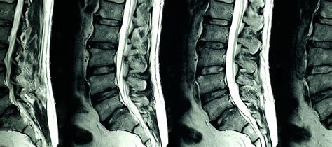
The lumbar spine, comprising the lower five vertebrae of the spinal column, is a complex anatomical structure that plays a crucial role in supporting the body’s weight, facilitating movement, and protecting the spinal cord. Magnetic Resonance Imaging (MRI) has become a cornerstone in the diagnosis and management of lumbar spine disorders, offering unparalleled soft tissue contrast and detailed visualization of the spinal anatomy. This guide aims to provide a comprehensive overview of the role of MRI in achieving accurate diagnoses of lumbar spine conditions, exploring its applications, interpretations, and the importance of integrating clinical correlation with imaging findings.
Introduction to Lumbar Spine MRI
Lumbar spine MRI is a non-invasive, painless procedure that utilizes a strong magnetic field and radio waves to generate detailed images of the lumbar spine. It is particularly useful for assessing the intervertebral discs, nerve roots, spinal cord, and surrounding soft tissues. The procedure is categorized based on the sequences used, including T1-weighted (T1W), T2-weighted (T2W), and Short-Tau Inversion Recovery (STIR) sequences, each providing unique information about the anatomical structures and potential pathologies.
Applications of Lumbar Spine MRI
- Disc Herniation: MRI is highly sensitive for detecting disc herniations, which occur when the gel-like nucleus pulposus protrudes through a tear in the outer, fibrous annulus. Herniated discs can compress adjacent nerve roots, leading to pain, numbness, and weakness in the lower extremities.
- Degenerative Disc Disease: This condition involves the gradual deterioration of the intervertebral discs, often associated with aging. MRI can assess the degree of disc dehydration, bulging, and any associated foraminal or central canal stenosis.
- Spinal Stenosis: This refers to the narrowing of the spinal canal, which can be congenital or acquired due to degenerative changes, tumors, or trauma. MRI can accurately measure the degree of stenosis and its impact on the spinal cord and nerve roots.
- Spondylolisthesis: This condition involves the anterior or posterior displacement of a vertebra, relative to the vertebra below. MRI can assess the degree of slippage and any associated nerve root compression or spinal cord injury.
- Infections and Inflammations: MRI is valuable in diagnosing infections such as discitis or osteomyelitis, as well as inflammatory conditions like ankylosing spondylitis, by demonstrating changes in the marrow signal and soft tissue involvement.
Interpreting Lumbar Spine MRI
Interpretation of lumbar spine MRI requires a systematic approach, focusing on the following aspects:
- Alignment: Assess the normal lordotic curvature of the lumbar spine and look for any signs of spondylolisthesis or scoliosis.
- Discs: Evaluate the signal intensity of the intervertebral discs on T2W images, noting any signs of dehydration (dark signal), herniation, or bulging.
- Nerve Roots and Spinal Cord: Look for compression, edema, or other abnormalities affecting these structures.
- Facets and Ligaments: Assess for signs of degenerative changes, such as facet joint hypertrophy or ligamentum flavum thickening, which can contribute to spinal stenosis.
- Soft Tissues: Evaluate the paraspinal muscles for signs of atrophy or strain and assess for any abnormal soft tissue masses.
Clinical Correlation
While MRI provides invaluable diagnostic information, it must be interpreted in the context of the patient’s clinical presentation. Symptoms, physical examination findings, and the results of other diagnostic tests (e.g., electromyography) are crucial for correlating MRI findings with the patient’s condition. For instance, a disc herniation evident on MRI may not necessarily be symptomatic, and the decision for surgical intervention often depends on the severity of symptoms and the failure of conservative treatments.
Future Directions
Advances in MRI technology continue to enhance the diagnostic capability for lumbar spine disorders. High-field strength MRI systems (3.0 Tesla and above) offer improved spatial resolution and better visualization of small structures. Furthermore, functional MRI techniques, such as diffusion tensor imaging, may provide insights into the microstructural integrity of the spinal cord and nerves, potentially guiding more personalized treatment strategies.
Conclusion
Lumbar spine MRI is a powerful diagnostic tool, offering detailed insights into the anatomy and pathology of the lower back. Its applications span a wide range of conditions, from degenerative diseases and disc herniations to infections and inflammatory conditions. However, the accurate diagnosis and management of lumbar spine disorders require a holistic approach, integrating MRI findings with clinical presentation, physical examination, and other diagnostic tests. As MRI technology continues to evolve, its role in guiding the treatment of lumbar spine conditions is likely to expand, contributing to better patient outcomes and quality of life.
FAQ Section
What are the primary uses of lumbar spine MRI in clinical practice?
+Lumbar spine MRI is primarily used for diagnosing conditions such as disc herniation, degenerative disc disease, spinal stenosis, spondylolisthesis, and infections or inflammatory diseases affecting the lumbar spine.
How does the interpretation of lumbar spine MRI differ from other types of spine MRI?
+While the fundamental principles of interpreting lumbar spine MRI are similar to those for other regions of the spine, the lumbar spine's unique anatomy and common pathologies (e.g., disc degeneration, facet joint osteoarthritis) require focused attention. The interpreter must be adept at recognizing the subtle signs of pathology that are specific to the lumbar region.
Can lumbar spine MRI be used as a screening tool for asymptomatic individuals?
+No, lumbar spine MRI is not recommended as a screening tool for asymptomatic individuals. The presence of asymptomatic disc bulges or degeneration is common, and such findings can lead to unnecessary concern or interventions. MRI should be reserved for individuals with significant symptoms or clinical findings suggestive of lumbar spine pathology.
Step-by-Step Guide to Preparing for a Lumbar Spine MRI
- Clothing: Wear comfortable, loose-fitting clothing without metal parts, as these can interfere with the MRI machine.
- Jewelry and Accessories: Remove all jewelry, including piercing, and avoid wearing clothes with metal buttons or zippers.
- Electronic Devices: Leave all electronic devices, such as smartphones, watches, and credit cards, outside the MRI suite.
- Claustrophobia: Inform your healthcare provider if you have claustrophobia, as arrangements can be made to make you more comfortable during the procedure.
- Medications: Continue to take your prescribed medications unless your doctor advises otherwise.
- Contrast Agents: In some cases, a contrast agent may be administered to enhance the visibility of certain structures. Be sure to inform your healthcare provider about any allergies or kidney problems.
By understanding the role of lumbar spine MRI in diagnosis and following the appropriate preparation steps, patients can better navigate their journey towards accurate diagnosis and effective treatment of lumbar spine conditions.