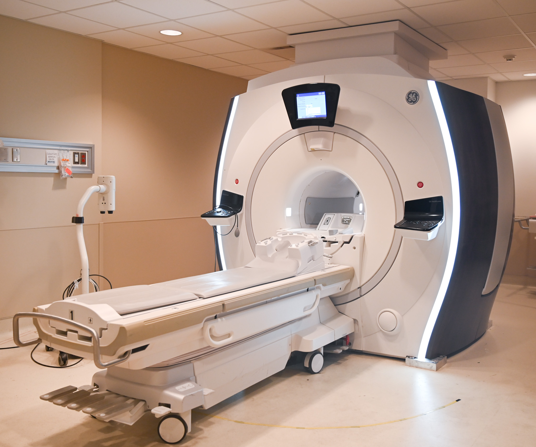
Mri Of L/S Spine: Comprehensive Guide

The lower spine, also known as the lumbar and sacral spine (L/S spine), is a complex region that plays a crucial role in supporting the body’s weight, facilitating movement, and protecting the spinal cord. Magnetic Resonance Imaging (MRI) of the L/S spine is a diagnostic imaging technique that provides detailed images of the spine, helping healthcare professionals to diagnose and treat various spinal conditions. In this comprehensive guide, we will delve into the world of MRI of the L/S spine, exploring its principles, applications, and interpretations.
Introduction to MRI of the L/S Spine
MRI is a non-invasive imaging modality that uses a strong magnetic field and radio waves to generate detailed images of the internal structures of the body. When it comes to the L/S spine, MRI is particularly useful for visualizing the soft tissues, including the spinal cord, nerve roots, and intervertebral discs. The L/S spine is comprised of five lumbar vertebrae (L1-L5) and five sacral vertebrae (S1-S5), which are fused together to form the sacrum.
Indications for MRI of the L/S Spine
MRI of the L/S spine is indicated for a wide range of conditions, including:
- Low back pain: MRI can help diagnose the underlying cause of low back pain, such as herniated discs, spinal stenosis, or spondylolisthesis.
- Sciatica: MRI can help identify the compression of nerve roots, which can cause sciatica symptoms.
- Spondylosis: MRI can help diagnose spondylosis, a condition characterized by the degeneration of the intervertebral discs.
- Spinal trauma: MRI can help evaluate the extent of spinal injuries, such as fractures or ligamentous injuries.
MRI Techniques for the L/S Spine
There are several MRI techniques that can be used to image the L/S spine, including:
- T1-weighted imaging: This technique provides detailed images of the spinal cord and nerve roots.
- T2-weighted imaging: This technique highlights the intervertebral discs and soft tissues.
- STIR (Short-Tau Inversion Recovery) imaging: This technique is useful for detecting bone marrow edema and inflammation.
- Gadolinium-enhanced imaging: This technique involves the use of a contrast agent to enhance the visibility of certain tissues.
Interpretation of MRI Images of the L/S Spine
Interpreting MRI images of the L/S spine requires a thorough understanding of spinal anatomy and pathology. The following structures should be evaluated:
- Spinal cord: The spinal cord should be evaluated for signs of compression, inflammation, or injury.
- Nerve roots: The nerve roots should be evaluated for signs of compression or injury.
- Intervertebral discs: The intervertebral discs should be evaluated for signs of degeneration, herniation, or infection.
- Vertebrae: The vertebrae should be evaluated for signs of fracture, osteoporosis, or tumor.
Common MRI Findings in the L/S Spine
Some common MRI findings in the L/S spine include:
- Herniated discs: A herniated disc can cause compression of the spinal cord or nerve roots.
- Spinal stenosis: Spinal stenosis can cause compression of the spinal cord or nerve roots.
- Spondylolisthesis: Spondylolisthesis can cause compression of the spinal cord or nerve roots.
- Degenerative disc disease: Degenerative disc disease can cause back pain and stiffness.
Clinical Applications of MRI of the L/S Spine
MRI of the L/S spine has several clinical applications, including:
- Diagnosis: MRI can help diagnose a wide range of spinal conditions.
- Treatment planning: MRI can help healthcare professionals develop treatment plans for spinal conditions.
- Monitoring: MRI can be used to monitor the progression of spinal conditions or the effectiveness of treatment.
Limitations and Challenges of MRI of the L/S Spine
While MRI of the L/S spine is a powerful diagnostic tool, it has several limitations and challenges, including:
- Cost: MRI is a relatively expensive imaging modality.
- Availability: MRI may not be available in all healthcare settings.
- Claustrophobia: Some patients may experience claustrophobia during the MRI procedure.
- Motion artifacts: Motion artifacts can degrade the quality of MRI images.
Future Directions of MRI of the L/S Spine
The field of MRI of the L/S spine is constantly evolving, with new techniques and applications being developed. Some future directions include:
- Advanced imaging techniques: New imaging techniques, such as diffusion tensor imaging and magnetic resonance spectroscopy, are being developed to provide more detailed information about spinal tissues.
- Artificial intelligence: Artificial intelligence algorithms are being developed to help healthcare professionals interpret MRI images more accurately and efficiently.
- Personalized medicine: MRI of the L/S spine can be used to develop personalized treatment plans for patients with spinal conditions.
What is the most common indication for MRI of the L/S spine?
+The most common indication for MRI of the L/S spine is low back pain. MRI can help diagnose the underlying cause of low back pain, such as herniated discs, spinal stenosis, or spondylolisthesis.
What is the difference between T1-weighted and T2-weighted imaging?
+T1-weighted imaging provides detailed images of the spinal cord and nerve roots, while T2-weighted imaging highlights the intervertebral discs and soft tissues.
Can MRI of the L/S spine help diagnose spinal trauma?
+Yes, MRI of the L/S spine can help evaluate the extent of spinal injuries, such as fractures or ligamentous injuries.
In conclusion, MRI of the L/S spine is a powerful diagnostic tool that provides detailed images of the spine, helping healthcare professionals to diagnose and treat various spinal conditions. While it has several limitations and challenges, the field of MRI of the L/S spine is constantly evolving, with new techniques and applications being developed. By understanding the principles, applications, and interpretations of MRI of the L/S spine, healthcare professionals can provide better care for patients with spinal conditions.


