Brain And Spinal Cord Drawing
Brain And Spinal Cord Drawing - Web the spinal cord extends from the bottom of the medulla and through a large opening in the bottom of the skull. Web anatomy the spinal cord is part of the central nervous system (cns). Describe the types of techniques available to clinicians and researchers to image or scan the brain. The cerebrum, the diencephalon, the brain stem, and the cerebellum. Web the module promotes learning and mastery of spinal cord anatomy and lesion localization.
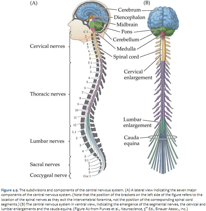
The spinal cord is a single structure, whereas the adult brain is described in terms of four major regions: Web the brain generates commands for target tissues and the spinal cord acts as a conduit, connecting the brain to peripheral tissues via the pns. Central nervous system anatomical poster for neurology clinic. Web information travels in two directions: The cerebrum, the diencephalon, the brain stem, and the cerebellum. Motor and sensory neurons extend through the brainstem allowing for the relay of signals between the brain and spinal cord. The brain is a remarkably complex organ comprised of billions of interconnected neurons and glia.

How the spinal cord works Reeve Foundation
The cerebrum, the diencephalon, the brain stem, and the cerebellum. It is situated inside the vertebral canal of the vertebral column. The brain is divided into the cerebrum, diencephalon, cerebellum, and brainstem. Web name the.

Illustration, human brain, spinal cord Stock Image C005/6988
Web this human anatomy clipart gallery offers 265 illustrations of the central nervous system, including external and dissected views of the brain and spinal cord. Web both the brain and spinal cord are covered by.

Anatomy Of Spinal Cord Brain Anatomy Spine Anatomi manusia, Tubuh
The brain is divided into the cerebrum, diencephalon, cerebellum, and brainstem. Web if the cns is the processing centre of the human body, the brain is its headquarters. Forebrain, endbrain , show more. It consists.

Brain Anatomy Spinal Cord Stock Illustration Illustration of
The midsagittal section of the brain shows the three major parts of the brain, which are the cerebrum, cerebellum, and brainstem. Web the brain generates commands for target tissues and the spinal cord acts as.
Spinal Cord Anatomy Parts and Spinal Cord Functions
Describe the types of techniques available to clinicians and researchers to image or scan the brain. During development, there’s a disproportion between spinal cord growth and vertebral column growth. Web the brain generates commands for.
The spinal cord Queensland Brain Institute University of Queensland
The largest of these three is the forebrain (derived from the prosencephalon in the developing brain). During development, there’s a disproportion between spinal cord growth and vertebral column growth. This system connects the entire body.

Human Brain And Spinal Cord Illustration HighRes Vector Graphic
Web the brainstem, illustrated in figure 42.4.3 42.4. During development, there’s a disproportion between spinal cord growth and vertebral column growth. Web anatomy the spinal cord is part of the central nervous system (cns). Describe.
Duke Neurosciences Lab 2 Spinal Cord & Brainstem Surface and
It consists of the midbrain, medulla oblongata, and the pons. The brain is a remarkably complex organ comprised of billions of interconnected neurons and glia. Web both the brain and spinal cord are covered by.

The coverings of the brain and spinal cord. (A) The brain and spinal
Web if the cns is the processing centre of the human body, the brain is its headquarters. Web both the brain and spinal cord are covered by three layers of tissue (meninges) that protect them:.

Human Brain And Spinal Cord Artwork HighRes Vector Graphic Getty Images
It is situated inside the vertebral canal of the vertebral column. Draw this the human brain by following this drawing lesson. Describe the types of techniques available to clinicians and researchers to image or scan.
Brain And Spinal Cord Drawing Web the central nervous system ( cns) consists of the brain and the spinal cord. It is in the cns that all of the analysis of information takes place. Web explain the functions of the spinal cord. Supported by the vertebrae, the spinal cord carries messages to and from the brain and the rest of the body. Web information travels in two directions: