Draw And Label The Human Cell
Draw And Label The Human Cell - They coordinate and function efficiently for the normal functioning of the cell. Cells walls should be visible: In humans, the haploid cells made in meiosis are sperm and eggs. Thin layer of protein and fat that surrounds the cell is the cell membrane. Web it’s time to label the cell yourself!
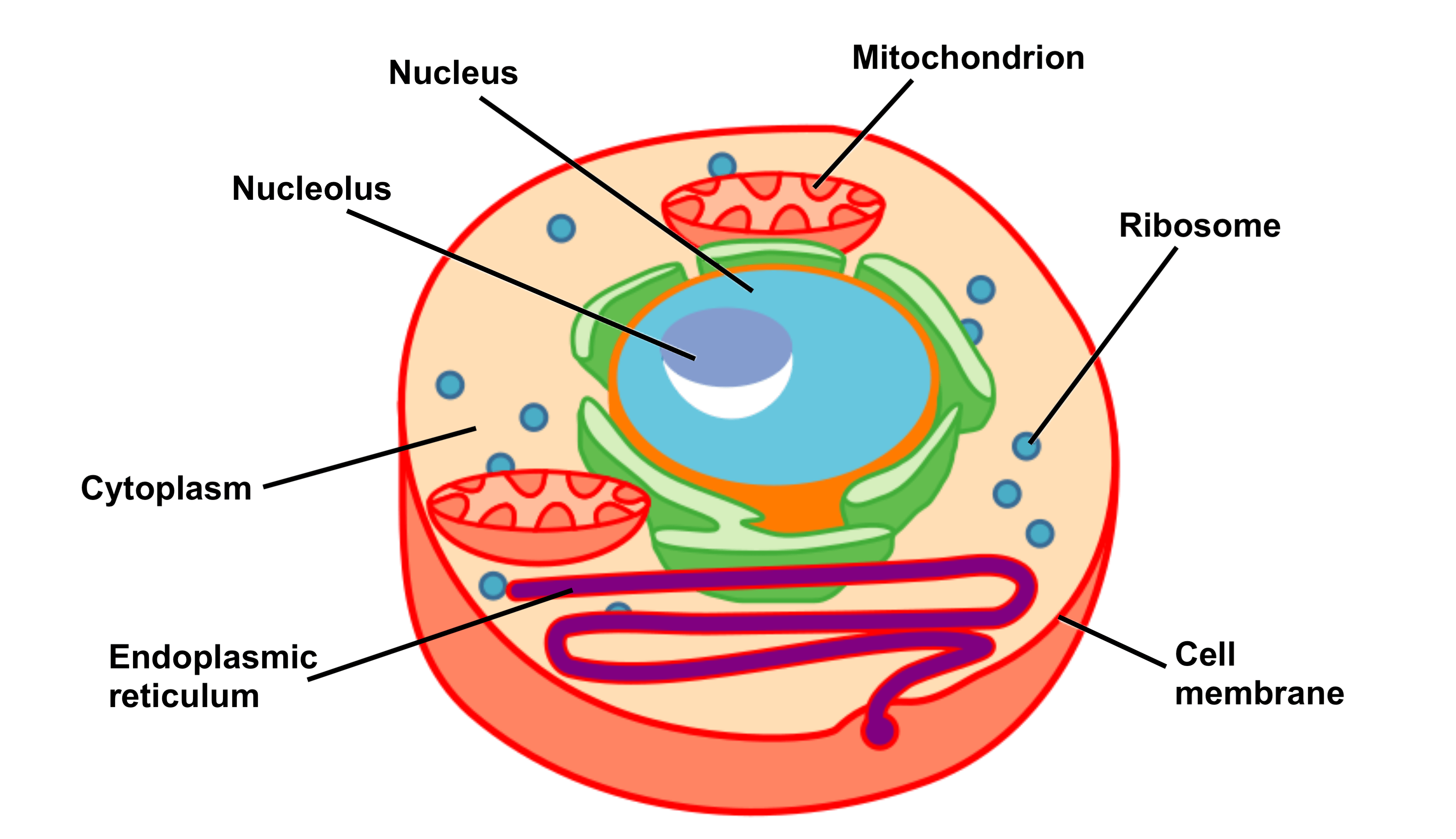
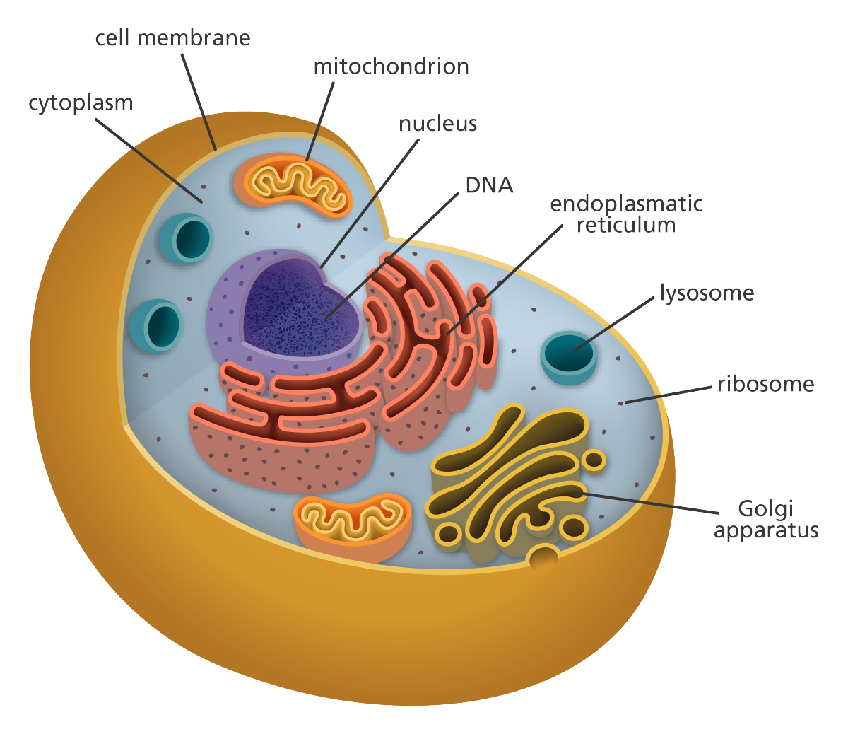
.§×hg$í \eqk‹)yi¢ ûqmñôoü] ’°a€ jb ûß›zï†ÿ{y[¹|$eêörî$±óçþûþêu7zj€d rj w¶šàÿ çtqöü÷ú ñ. Web to put that another way, meiosis in humans is a division process that takes us from a diploid cell—one with two sets of chromosomes—to haploid cells—ones with a single set of chromosomes. A single cell is often a complete organism in itself, such as a bacterium or yeast. Draw various components of the cell shapes in long thin lines. How many nucleoli are present in each nucleus? Web hey guys!!!in this drawing,i will show you how to draw and label a simple human cell easy step by step for beginnersin this video, i have used : The outermost part of the cell, which is shown as a thick outline of the figure, is labeled cell wall.

Human Cell Diagrams Human cell diagram, Physiology, Anatomy and
In other words, it is the series of growth and development steps a cell undergoes between its “birth”—formation by the division of a mother cell—and reproduction—division to make two new daughter cells. Web structure and.
Human Cell Sketch at Explore collection of Human
The membrane, the nucleus, and the. Highlight the inner portion of the cell known as cytoplasm with a yellow marker. Then you’ll draw a bigger circle around that. Web a cell is the smallest living.
Learn the parts of a cell with diagrams and cell quizzes Kenhub
Web it’s time to label the cell yourself! Make the individual cells 20 mm wide. A group of cells forms tissue, various tissues forms an organ and different organs make up the body. For a.
Human cell diagram Etsy
A group of cells forms tissue, various tissues forms an organ and different organs make up the body. Web there are many different types, sizes, and shapes of cells in the body. They coordinate and.

How to Draw Human Cell Step by Step YouTube
Gametes are sex cells that join together during sexual reproduction. Sketch the cell at low and high power. Begin by making a big circle and lines connecting it to label the cells. Web you can.
Education Chart of Biology for Human Cell Diagram Best Acupuncture llc
Then, where an animal cell would go through cytokineses, a plant cell simply creates a new cell plate in the middle, creating two new cells. Somatic cells are all the cells in the body that.
Human Cell Diagram 6406474 Vector Art at Vecteezy
This bigger circle is the nucleus. The cell plate later changes to a cell wall once the division is complete. Gametes are sex cells that join together during sexual reproduction. Nucleus, nucleoli, nuclear envelope, cytoplasm,.
Cell Structure and Function Part 1 The Organelles Medical Exam Prep
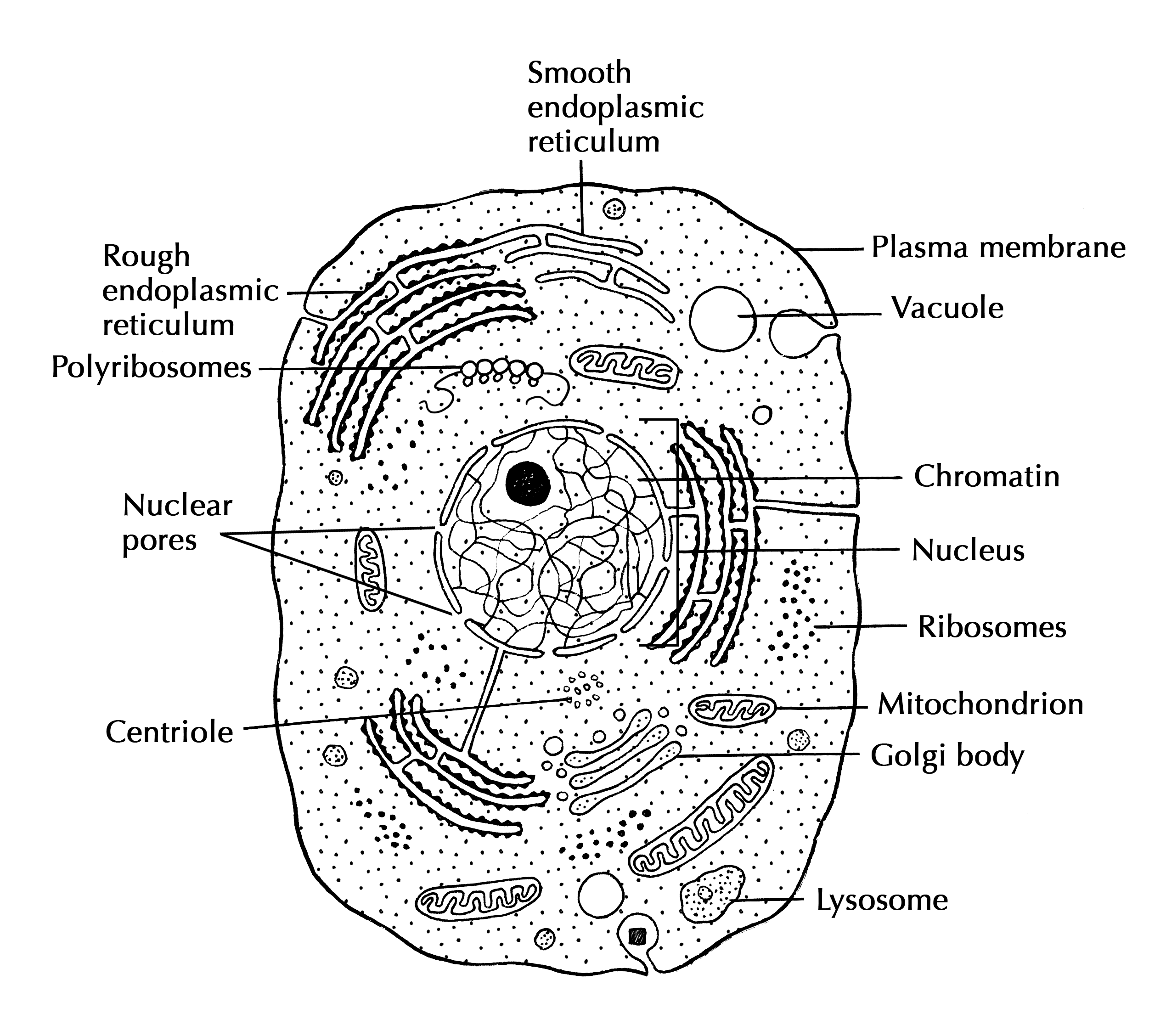
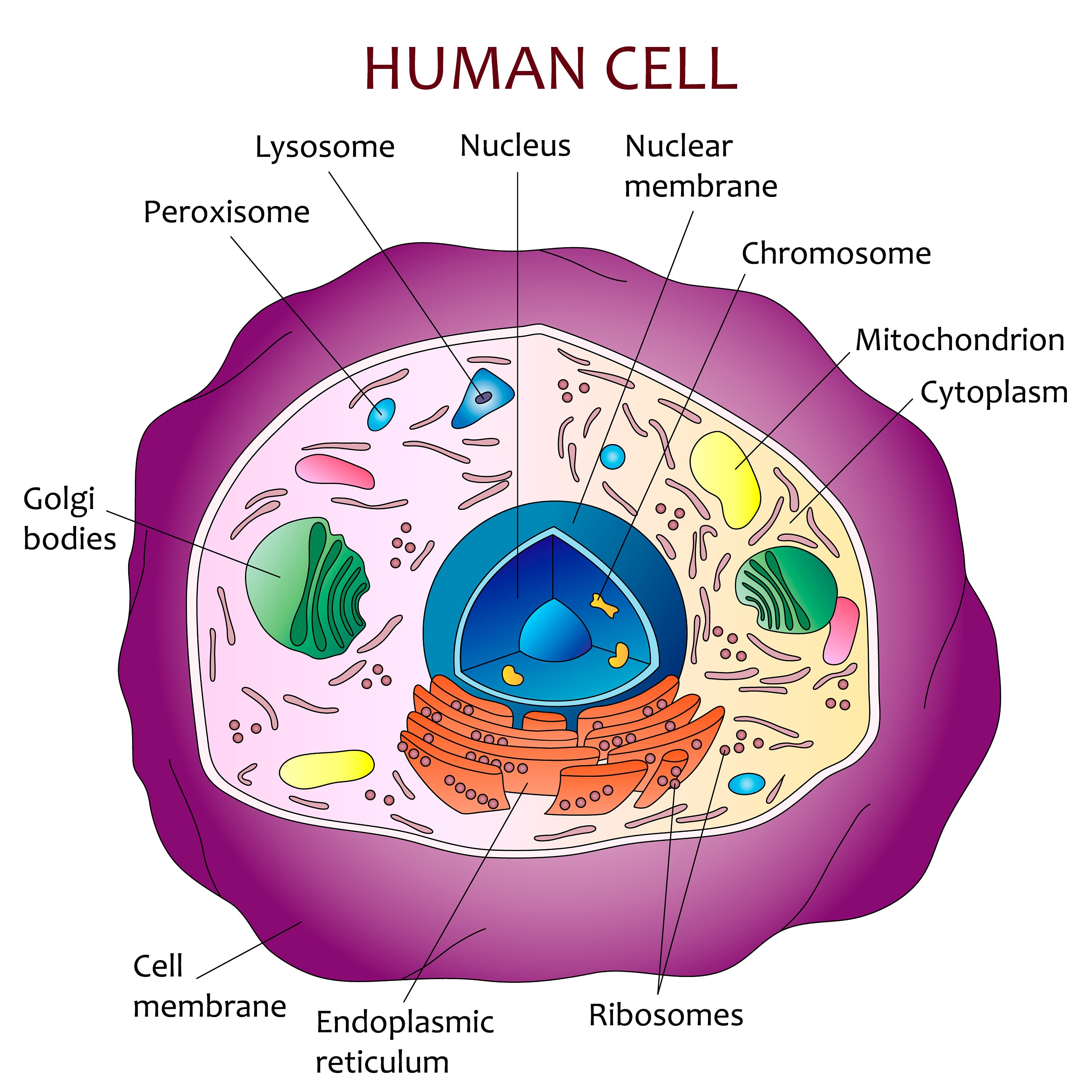
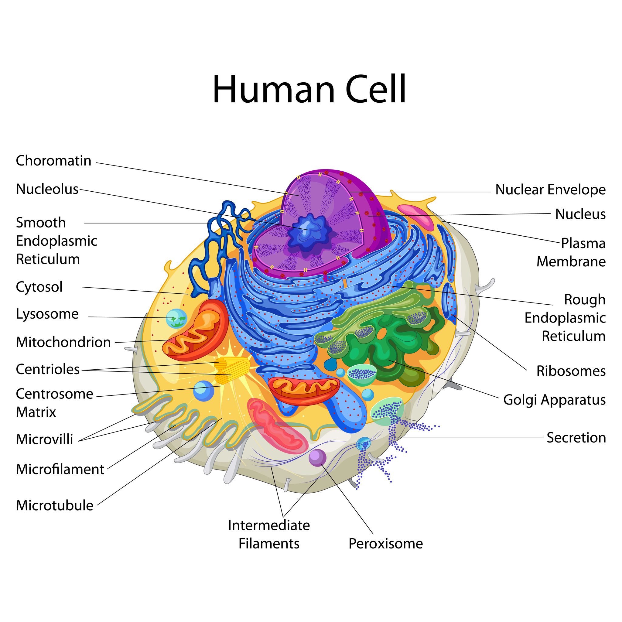
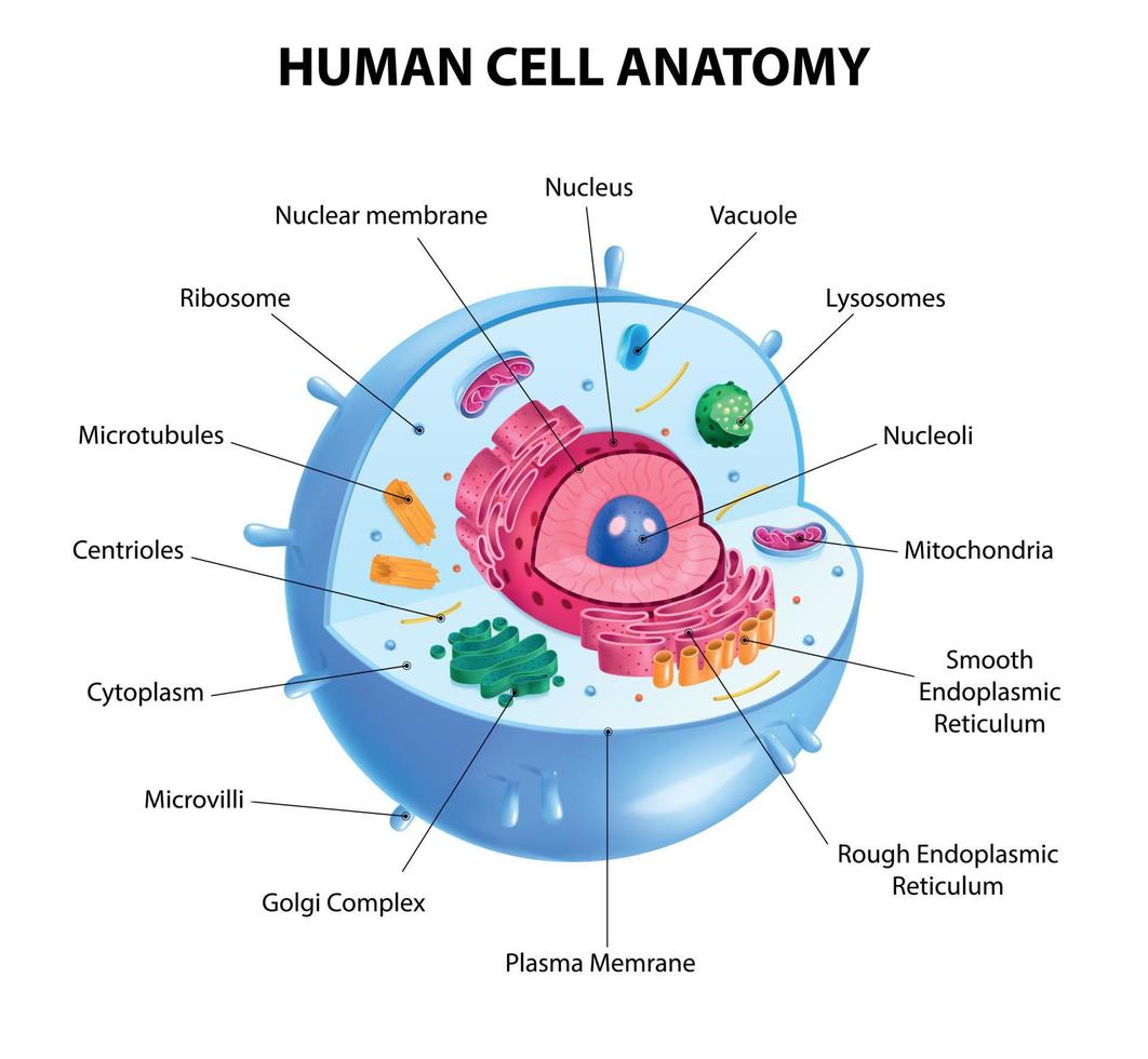
How many nucleoli are present in each nucleus? Thin layer of protein and fat that surrounds the cell is the cell membrane. The structure and components of a human cell are given below: Web the.
The Cell Theory & Structure HubPages
Web there are hundreds of different types of cells in the human body, which vary in shape (e.g. Begin by making a big circle and lines connecting it to label the cells. (interphase, prophase, metaphase,.

Human Cell Diagram, Parts, Pictures, Structure and Functions
Web there are many different types, sizes, and shapes of cells in the body. The outermost part of the cell, which is shown as a thick outline of the figure, is labeled cell wall. For.
Draw And Label The Human Cell (interphase, prophase, metaphase, anaphase, telophase). Together, trillions of cells make up the human body. Small granule cells of the cerebellum in the brain (4 micrometers), up to the huge oocytes (eggs) produced in the female reproductive organs (100 micrometers) and function. As you fill in the cell structure worksheet, remember the functions of each part of the cell that you learned in the video. The structure and components of a human cell are given below: