Draw And Label The Thoracic Vertebrae
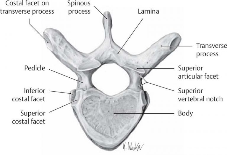
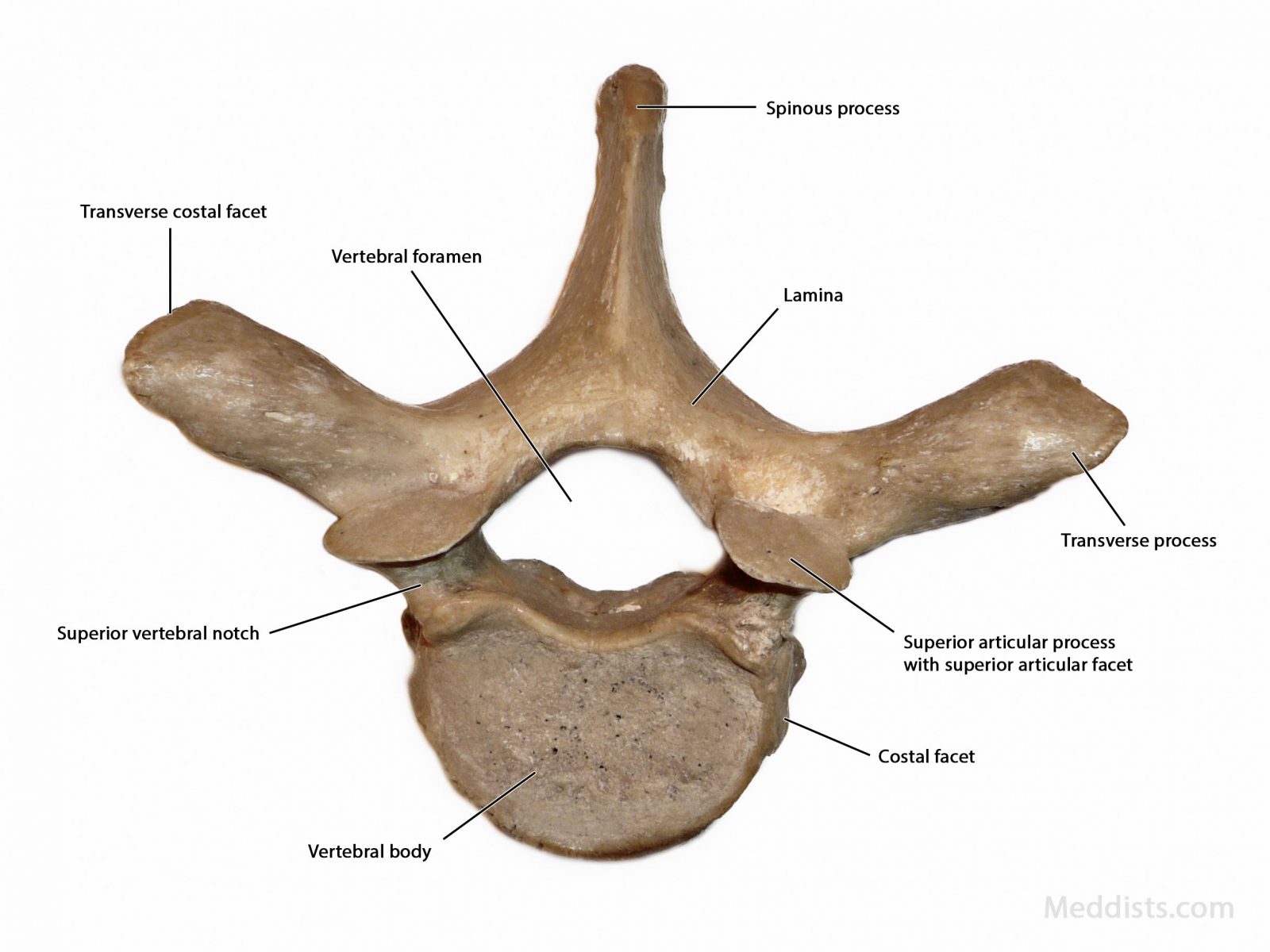
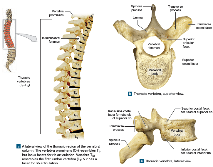
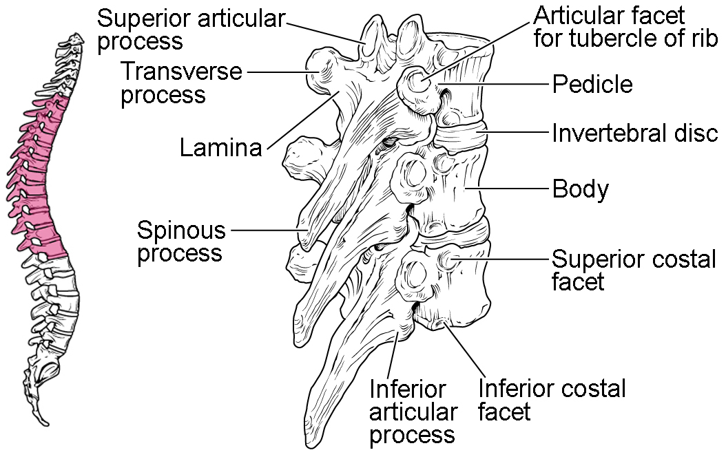
Draw And Label The Thoracic Vertebrae - The twelve thoracic vertebrae are strong bones that are located in the middle of the vertebral column, sandwhiched between the cervical ones above and the lumbar vertebrae below. They increase in size going towards the lumbar vertebrae, with the lower ones being much larger than the upper. 12 thoracic vertebrae, located in the upper area of the back. Web the thoracic vertebrae (latin: Web 1 2 vertebral arch or neural arch (a rcus vertebrae) is made up of two pedicles, two laminae, and a spinous process.
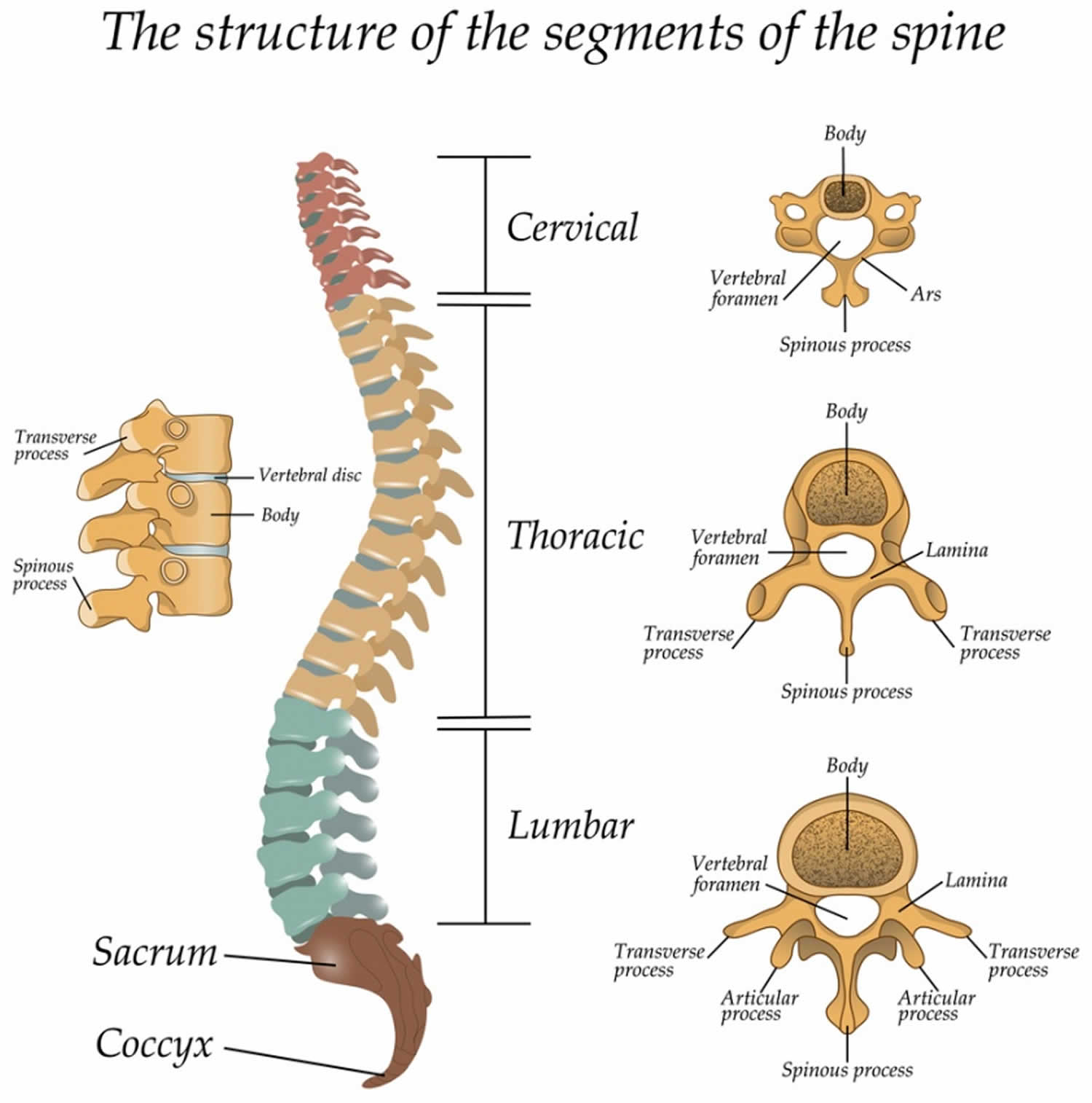
The lumbar vertebrae are intermediate in size and keep on increasing towards the lumbar vertebrae. Web moving forward with the skeletal scaffold of the thorax, we have the thoracic skeleton. This is an online quiz called label the thoracic vertebrae. Web in humans, there are twelve thoracic vertebrae and they are intermediate in size between the cervical and lumbar vertebrae; There is a printable worksheet available for download here so you can take the quiz with pen and paper. Typical cervical vertebrae, such as c4 or c5, have several characteristic features that differentiate them from thoracic or lumbar vertebrae (). Web the thoracic wall is bounded anteriorly by the sternum and costal cartilages;
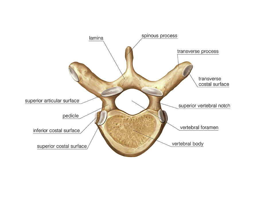
Thoracic vertebrae diagram
Web thoracic vertebrae have sites for rib attachment, and the vertebrae that give rise to the sacrum and coccyx have fused together into single bones. The bodies of the thoracic vertebrae are larger than those.
Thoracic Vertebra Photograph by Asklepios Medical Atlas Pixels
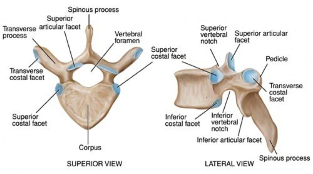
This article will look at the osteology of the thoracic vertebrae, examining their characteristic. You can use it as thoracic vertebrae quiz practice, completely free to play. These vertebrae are characterized by their articulation with.
Thoracic vertebrae anatomy, function & thoracic vertebrae injury
Web the thoracic vertebrae (latin: The manubrium, body, and xiphoid process. Web thoracic vertebrae have sites for rib attachment, and the vertebrae that give rise to the sacrum and coccyx have fused together into single.
Thoracic Vertebrae (Thoracic Spine) Anatomy & Labeled Diagram
Web thoracic vertebrae have sites for rib attachment, and the vertebrae that give rise to the sacrum and coccyx have fused together into single bones. There is a printable worksheet available for download here so.
Anatomy of the Thorax → Thoracic Vertebral Column
7 cervical vertebrae, which are located in the neck. You can use it as thoracic vertebrae quiz practice, completely free to play. You can use it as label the thoracic vertebrae practice, completely free to.
Thoracic vertebrae structure, function, Chest wall muscles
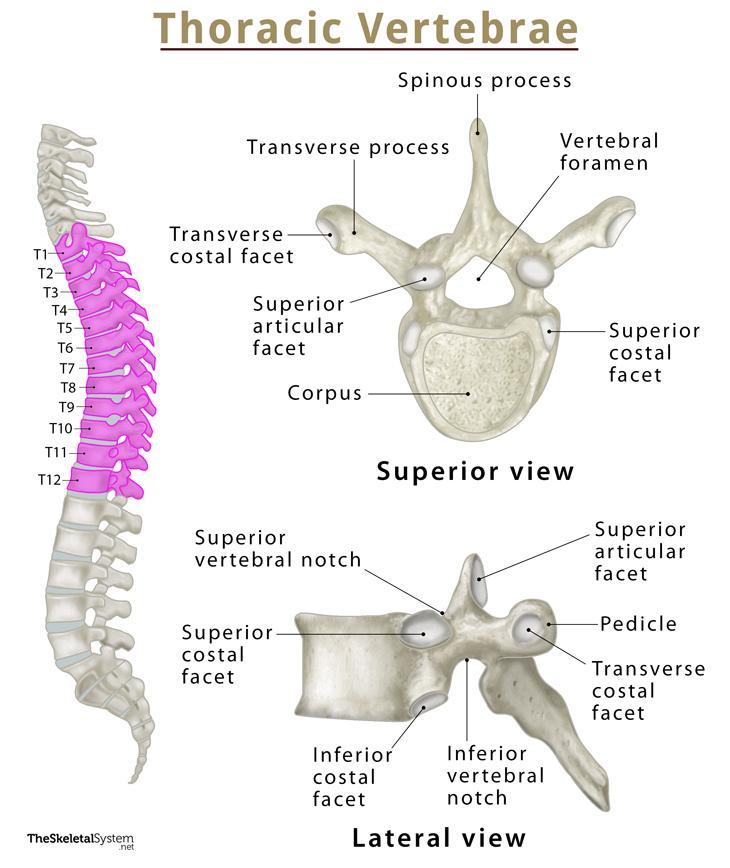
Vertebrae are the 33 individual, interlocking bones that form your spinal column. Your thoracic spine consists of 12 vertebrae, labeled t1 through t12. There are twelve in total (t1 to t12), which together form the.
Thoracic vertebrae anatomy, function & thoracic vertebrae injury
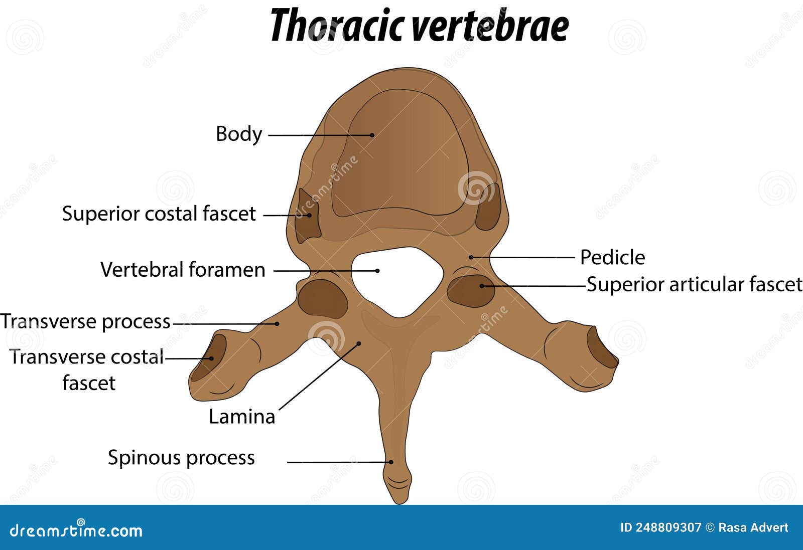
They are thicker and larger than the cervical vertebrae but smaller than the lumbar vertebral bones. Web 1 2 vertebral arch or neural arch (a rcus vertebrae) is made up of two pedicles, two laminae,.
Anatomy of the Thoracic Vertebrae Labeled Diagram Vector Illustration
Web the thoracic wall is bounded anteriorly by the sternum and costal cartilages; It is made up of the sternum, twelve pairs of ribs, twelve thoracic vertebrae, and interconnecting joints. Typical cervical vertebrae, such as.

diagram of thoracic vertebra
Superiorly by the suprapleural membrane and inferiorly by the respiratory diaphragm. These vertebrae are characterized by their articulation with the ribs. The manubrium, body, and xiphoid process. Web by definition, the vertebrae are the bones.
The Vertebral Column Anatomy and Physiology I
There are twelve in total (t1 to t12), which together form the thoracic spine. The main thoracic joints include the intervertebral discs, costovertebral, sternocostal, sternoclavicular, costochondral, and interchondral joints. Laterally by the ribs and intercostal.
Draw And Label The Thoracic Vertebrae Web thoracic vertebrae quiz — quiz information. Web in humans, there are twelve thoracic vertebrae and they are intermediate in size between the cervical and lumbar vertebrae; 12 thoracic vertebrae, located in the upper area of the back. Superior view of the vertebral arch of a vertebra. Web the thoracic vertebrae are bones located between the cervical and lumbar vertebrae.