Drawing Of Simple Squamous Epithelium
Drawing Of Simple Squamous Epithelium - Web distinguish between simple epithelia and stratified epithelia, as well as between squamous, cuboidal, and columnar epithelia describe the structure and function of endocrine and exocrine glands and their respective secretions • how to draw simple cuboidal. They are sketches from selected slides used in class from the teaching slide set. Try to identify the simple squamous epithelia in these pictures. Simple epithelium can be divided into 4 major classes, depending on the.
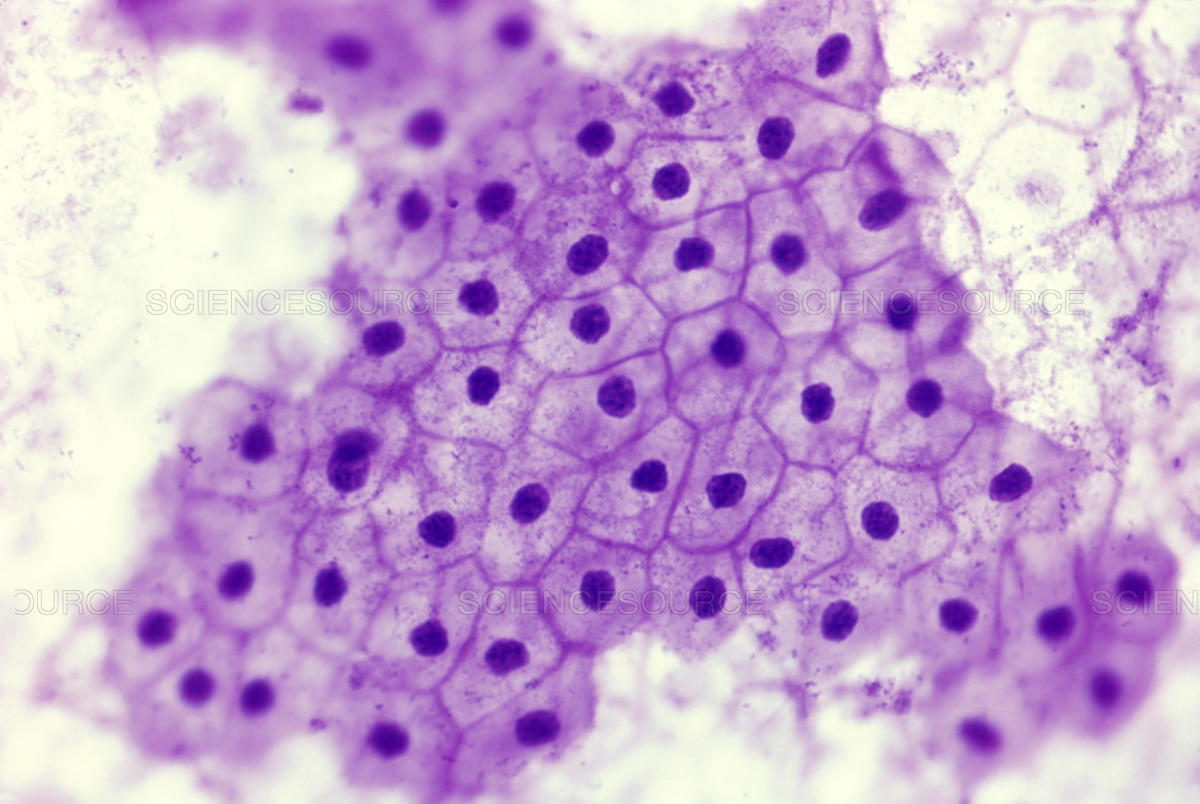
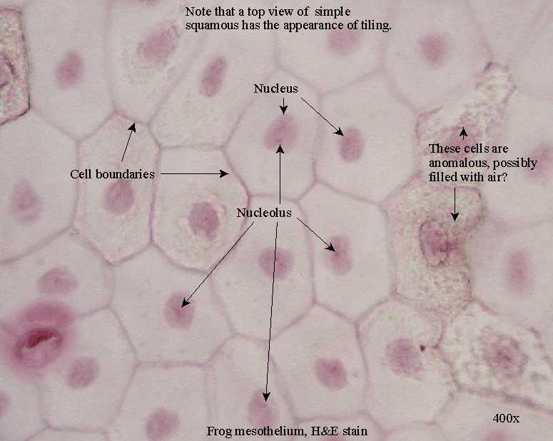
Try to identify the simple squamous epithelia in these pictures. The compound epithelium has two or more. These are the nuclei of the simple squamous epithelial cells. Simple epithelium can be divided into 4 major classes, depending on the. Cuboidal lines small ducts, tubules. The simple epithelium is a single layer of cells that line the ducts, body cavities and tubes. Web first, you should draw the basement membrane of the simple squamous epithelium.

simple squamous epithelium Google Search Histology slides, Squamous
Web epithelial tissue or epithelium are tightly packed cells that act as a barrier between the exterior and interior. Web distinguish between simple epithelia and stratified epithelia, as well as between squamous, cuboidal, and columnar.
Simple Squamous Epithelium Inrtroducrion , Anatomy & Function
They are sketches from selected slides used in class from the teaching slide set. Most of these cells arise from the ectoderm, or outermost layer of. The compound epithelium has two or more. Simple squamous.

Simple Squamous Epithelium Location And Function Steve Gallik
Web one of the problems with this type of tissue preparation is that the cells lose most of their connections to each other. • how to draw simple cuboidal. This is made up of thin,.
Epithelial Tissue Anatomy & Physiology
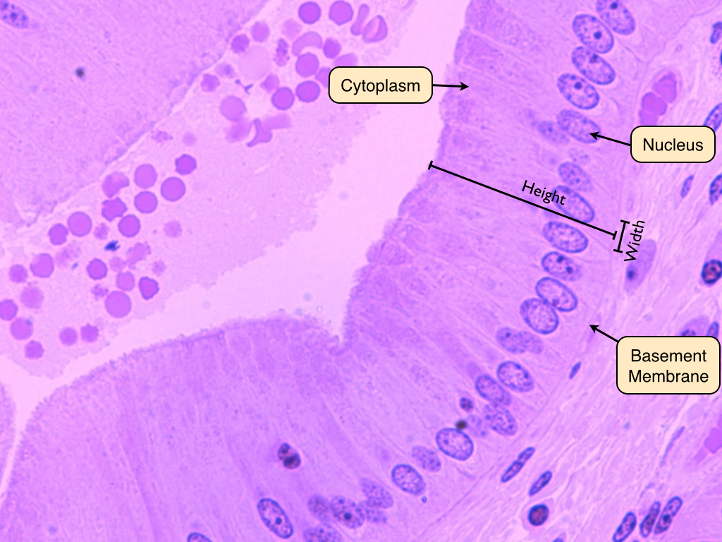
• how to draw simple squamous epitheliu. The basement membrane provides support to the endothelium or epithelium and the tissue. Web epithelial tissue or epithelium are tightly packed cells that act as a barrier between.

How to draw stratified squamous epithelium easy way YouTube
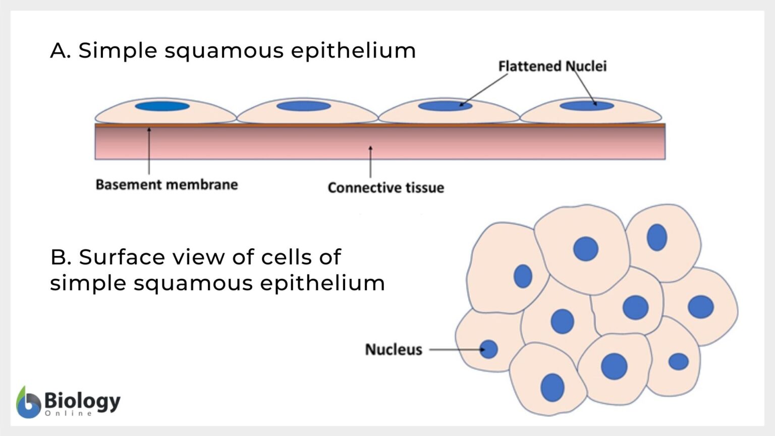
Web epithelial tissue or epithelium are tightly packed cells that act as a barrier between the exterior and interior. This is made up of thin, flat and hexagonal cells. The basement membrane provides support to.
Simple Squamous Epithelium Inrtroducrion , Anatomy & Function
Web distinguish between simple epithelia and stratified epithelia, as well as between squamous, cuboidal, and columnar epithelia describe the structure and function of endocrine and exocrine glands and their respective secretions Web simple squamous epithelium.
Simple squamous epithelium Definition and Examples Biology Online
Web hello friends, this is my youtube channel and in this channel i used to share videos of different diagrams in easy way and step by step tutorials. Both the endothelial lining of blood vessels.

Simple Squamous Epithelium Pseudostratified Columnar Epithelium Simple
Web the drawings of histology images were originally designed to complement the histology component of the first year medical course run prior to 2004. Try to identify the simple squamous epithelia in these pictures. Cuboidal.

Simple Squamous Epithelium Diagram Quizlet
Web one of the problems with this type of tissue preparation is that the cells lose most of their connections to each other. Simple squamous epithelia are found in a variety of locations, starting from.

Histology Image Membranous epithelium
They can be classified into simple and compound epithelium based on the number of cell layers. Simple columnar epithelium stratified squamous epithelium simple cuboidal epithelium transitional epithelium. A columnar epithelial cell looks like a column.
Drawing Of Simple Squamous Epithelium The alveoli of lungs where gases diffuse, segments of kidney tubules, and the lining of capillaries are also made of simple squamous epithelial tissue. Web first, you should draw the basement membrane of the simple squamous epithelium. Simple epithelium can be divided into 4 major classes, depending on the. A few epithelial layers are constructed from cells that are said to have a transitional shape. Simple squamous epithelia are found in a variety of locations, starting from capillaries to the alveoli of lungs, and nephrons of kidneys.