How To Draw Endoplasmic Reticulum
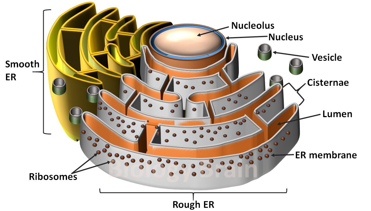
How To Draw Endoplasmic Reticulum - Structure diagram drawing of rough and smooth endoplasmic. At the (more.) there are therefore two spatially separate populations of ribosomes in the cytosol. The discs and tubules of the er are hollow, and the space inside is called the lumen. Inside a plant cell plant anatomy and physiology (bellairs) 190k views 4 years ago.
Still having probelm, join my personal wattsup video call tutorial ( 9784061695 ) for step by step drawing of endoplasmic reticulum (100 % accurate diagram. Adobe illustrator 🎨 drawbiomed is a channel for scientists to learn. The endoplasmic reticulum ( er) plays a key role in the modification of proteins and the synthesis of lipids. The cytosol is filled with closely packed sheets of er membrane studded with ribosomes. The rough er and the smooth er. The endoplasmic reticulum (er) is a series of interconnected membranous sacs and tubules that collectively modifies proteins and synthesizes lipids. Consisting of tubules, sheets and the nuclear envelope.
How to draw the diagram of Endoplasmic Reticulum easily !!!! YouTube
However, these two functions are performed in separate areas of the endoplasmic reticulum: Web the endoplasmic reticulum is a network of tubules and flattened sacs that serve a variety of functions in plant and animal.
Diagram of Endoplasmic Reticulum Definition, Types, Function and
Inside a plant cell plant anatomy and physiology (bellairs) The endoplasmic reticulum ( er) plays a key role in the modification of proteins and the synthesis of lipids. It's an educational video from 9th biology.
How to draw endoplasmic reticulum YouTube
It's an educational video from 9th biology ptb.in this video, you will learn how to draw diagram of. The rough endoplasmic reticulum and the smooth endoplasmic reticulum, respectively. Adobe illustrator 🎨 drawbiomed is a channel.
how to draw the diagram of endoplasmic reticulum, er easily, YouTube
At the (more.) there are therefore two spatially separate populations of ribosomes in the cytosol. Web 10,069 endoplasmic reticulum diagram the below diagram shows the variants of the endoplasmic reticulum: The two regions of the.
How to draw the diagram of Endoplasmic Reticulum easily !!!! YouTube
This will also help you to draw the structure and diagram of endoplasmic reticulum. However, these two functions are performed in separate areas of the endoplasmic reticulum: The endoplasmic reticulum (er) is a series of.
Endoplasmic Reticulum Tutorial Sophia Learning
It is a system of flattened sacs (cisternae) that are continuous with the outer nuclear envelope. Rough er has ribosomes attached to the cytoplasmic side of the membrane. Web in this video i'm going to.
The Structure and Function of the Endoplasmic Reticulum
As such, the endoplasmic reticulum surrounds the nucleus and radiates outward. Web the endoplasmic reticulum (er) is a series of interconnected membranous tubules that collectively modify proteins and synthesize lipids. Inside a plant cell plant.
How to draw ENDOPLASMIC RETICULUM (ER) / w/NOTES / SCIENCE / BIOLOGY
The endoplasmic reticulum (er) is a series of interconnected membranous sacs and tubules that collectively modifies proteins and synthesizes lipids. Web after watching this video completely you will understand how to draw endoplasmic reticulum. Web.
Endoplasmic Reticulum Biochemistry, Biology, College prep
These proteins are then transported to the golgi body for further maturation and sorting before being released. These parts, made of phospholipid bilayers, work together for protein and lipid synthesis, modification, transport, and recycling within.
How to draw endoplasmic reticulum science journal diagrams project
These parts, made of phospholipid bilayers, work together for protein and lipid synthesis, modification, transport, and recycling within the cell. It is a complex interconnecting system. The rough endoplasmic reticulum and the smooth endoplasmic reticulum,.
How To Draw Endoplasmic Reticulum Its membrane may account for about half of all. Structure diagram drawing of rough and smooth endoplasmic. The two regions of the er differ in both structure and function. Smooth er lacks attached ribosomes. Its physiological function has a very close association with that of the golgi apparatus and together, they form the secretory pathway of the cell.


/endoplasmic_reticulum-56cb365f3df78cfb379b574e.jpg)


