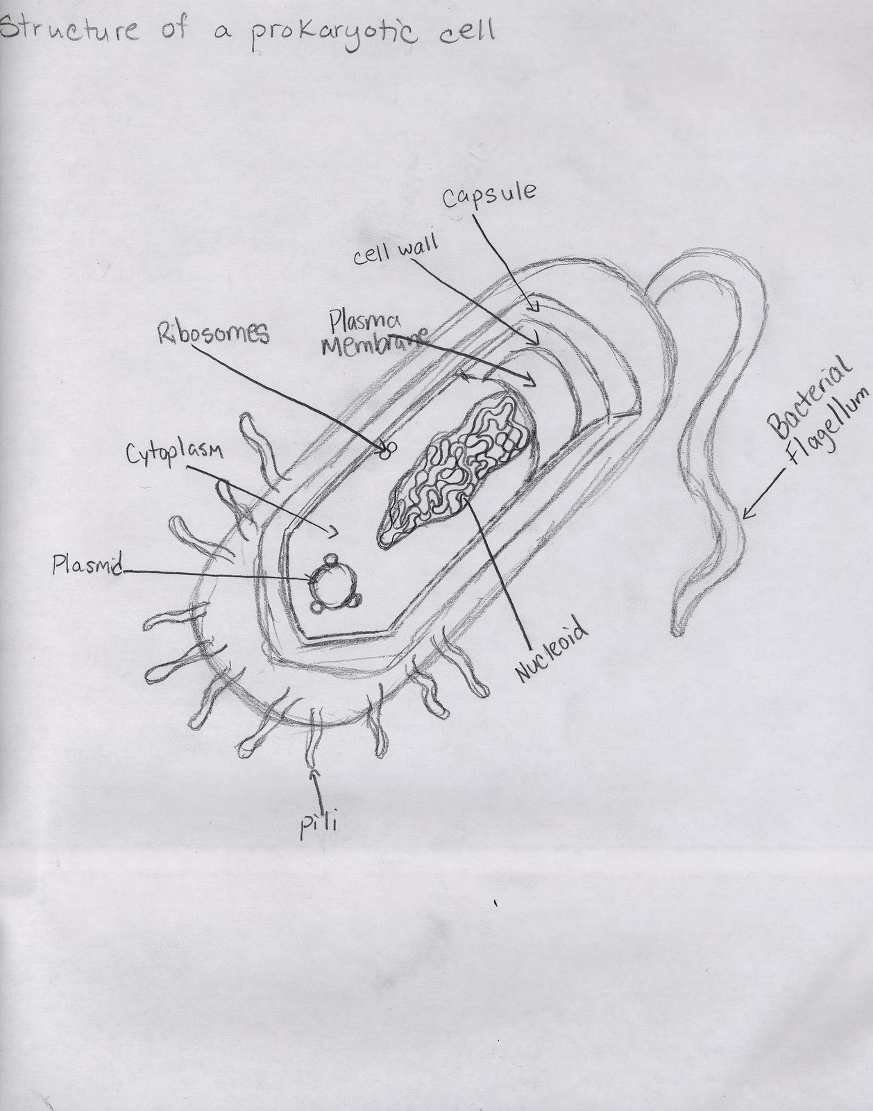
How To Draw Prokaryotic Cell
How To Draw Prokaryotic Cell - Web scientists believe that prokaryotic cells were some of the first life forms on earth. Describe the functions of the structures found in prokaryotic. Undergo mitosis (for somatic cells) or meiosis (for sex cells) during cell division. All cells share four key components: Many also have a capsule or slime layer made of polysaccharide.
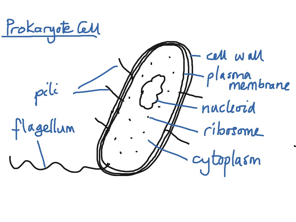
These neat, well labelled and. Some prokaryotes may have additional structures such as a capsule, flagella, and pili. The plasma membrane is an outer covering that separates the cell’s interior from its surrounding environment. Describe the functions of the structures found in prokaryotic. Web about press copyright contact us creators advertise developers terms privacy policy & safety how youtube works test new features nfl sunday ticket press copyright. Play this screencast how to draw a prokaryote cell for ib biology. All cells share four key components:
Prokaryotic Cell Drawing Prokaryotic Cells Simple Diagram Diagram
All prokaryotic cells are encased by a cell wall. Unlike mitosis, this process does not involve the condensation of dna or the duplication of organelles. Undergo mitosis (for somatic cells) or meiosis (for sex cells).
HOW TO DRAW A PROKARYOTIC CELL. YouTube
Web most prokaryotes have a cell wall outside the plasma membrane. Web about press copyright contact us creators advertise developers terms privacy policy & safety how youtube works test new features nfl sunday ticket press.
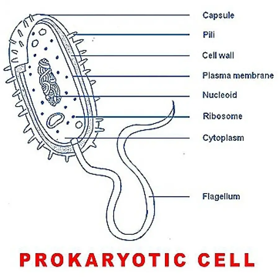
Draw a well labelled diagram of a prokaryotic cell Brainly.in
Reproduce through binary fission, where the cell splits into two identical daughter cells. The structure called a mesosome was once thought to be an organelle. Prokaryotic cells have only a small amount of dna, which.
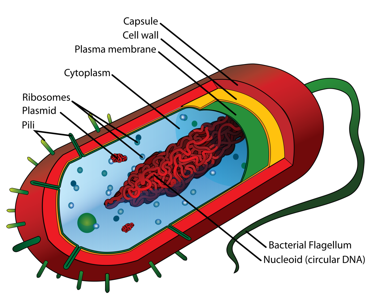
Prokaryotic Cell Structure A Visual Guide Owlcation
The composition of the cell wall differs significantly between the domains bacteria and archaea, the two domains of life into which prokaryotes are divided. Like other prokaryotic cells, this bacterial cell lacks a nucleus but.
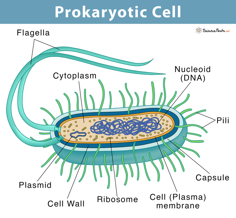
Prokaryotic Cell Definition, Examples, & Structure
A classic example of a prokaryotic cell is escherichia coli (e. Web most prokaryotes have a cell wall outside the plasma membrane. They have a single piece of circular dna in the nucleoid area of.
Draw a diagram of a prokaryotic cell and label at least four parts in it.
Some prokaryotes may have additional structures such as a capsule, flagella, and pili. The features of a typical prokaryotic cell are shown. The main parts of a prokaryotic cell are shown in this diagram. The.
How to draw a prokaryotic cell prokaryotic organism Bacterial cell
The plasma membrane is an outer covering that separates the cell’s interior from its surrounding environment. Many also have a capsule or slime layer made of polysaccharide. These neat, well labelled and. Play this screencast.
Simple Prokaryotic Cell Diagram
Web prokaryotic cells divide through the process of binary fission. The prokaryotic cell diagram given below represents a bacterial cell. Undergo mitosis (for somatic cells) or meiosis (for sex cells) during cell division. This diagram.
Cell Types and Structure Structure of Prokaryotic Cell
The plasma membrane is an outer covering that separates the cell’s interior from its surrounding environment. Like other prokaryotic cells, this bacterial cell lacks a nucleus but has other cell parts, including a plasma membrane,.
How to draw easily PROKARYOTIC CELLS / STRUCTURE and FUNCTION / w
Describe the functions of the structures found in prokaryotic. They have no true nucleus as the dna is not contained within a membrane or separated from the rest of the cell, but is coiled up.
How To Draw Prokaryotic Cell Web i draw a bacterial cell to show you how to make an accurate biological drawing of a prokaryotic cell. After you are finished with the slides, clean the immersion oil from the microscope lens. Identify each of these parts in the diagram. Prokaryotic cells are fundamental to mastering high school cell biology. Web in this activity, students will work in pairs to doodle two cell models, one of a prokaryotic cell, and one of a eukaryotic cell.

