Microscope Draw Tube
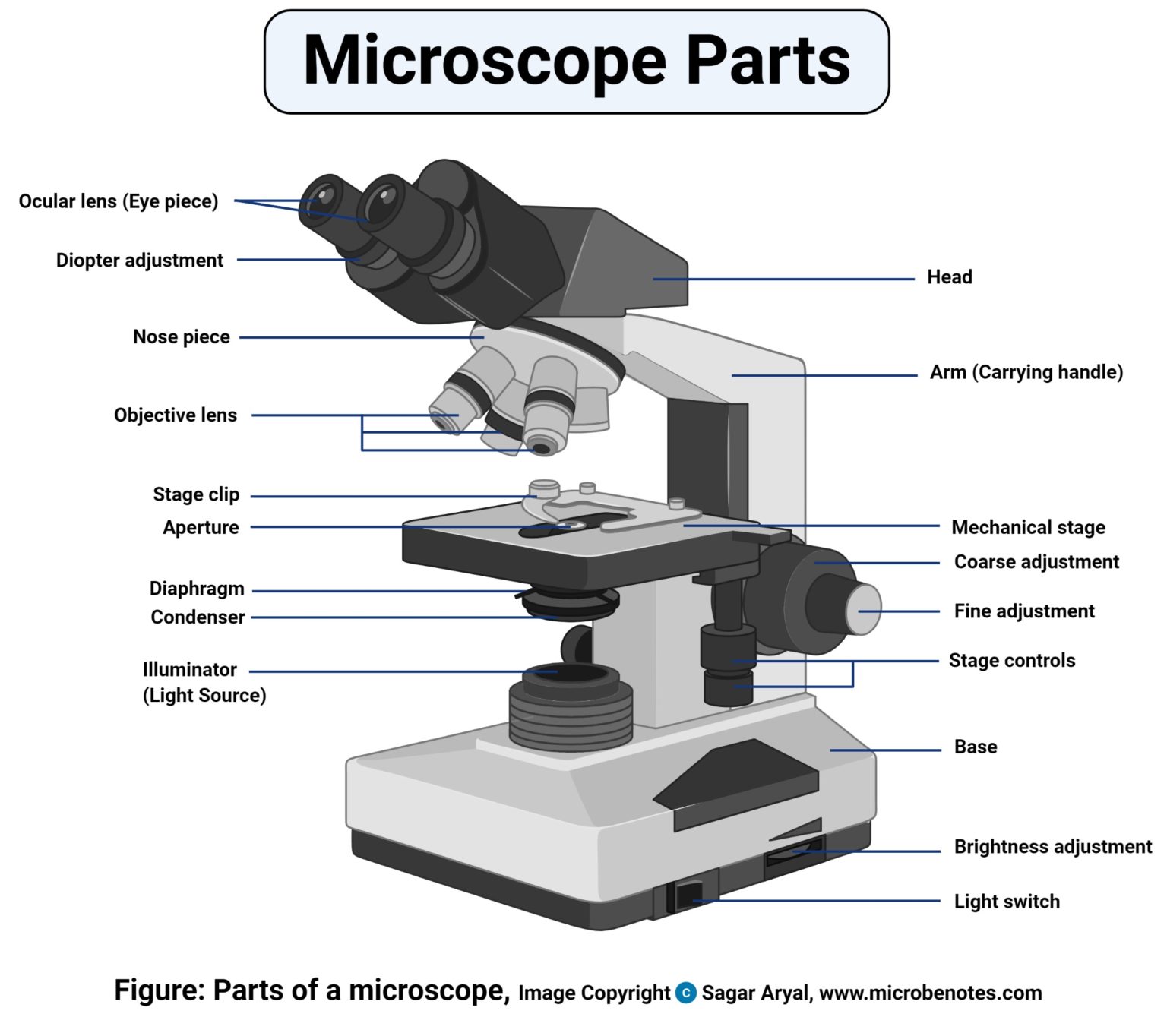
Microscope Draw Tube - Web connects ocular tube and base. Shape the microscope head 1.3 step 3: Web the mechanical tube length of an optical microscope is defined as the distance from the nosepiece opening, where the objective is mounted, to the top edge of the observation tubes where the eyepieces (oculars) are inserted. For the passage of light rays through the body tube, there is a pathway. Stage control/stage height adjustment 16.
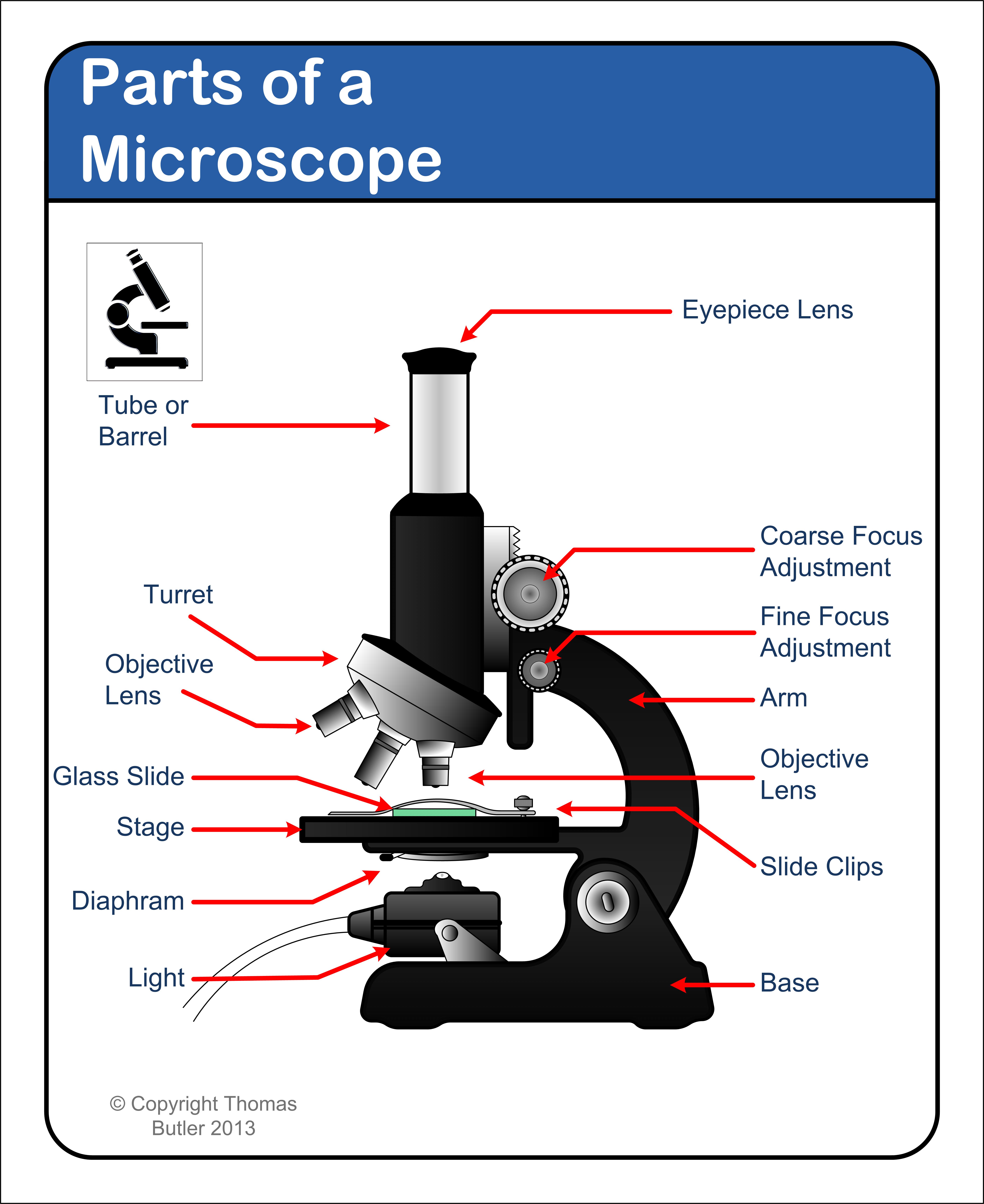
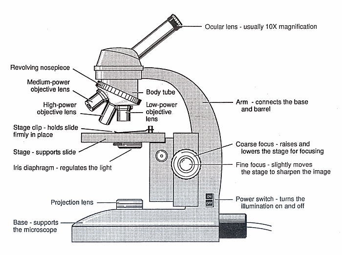
Provides support to help microscope stand upright: Web this short video discuss the expectations of a microscope observation and drawings and also provides examples of errors to watch out for.teachers: Web the mechanical tube length of an optical microscope is defined as the distance from the nosepiece opening, where the objective is mounted, to the top edge of the observation tubes where the eyepieces (oculars) are inserted. It is the tube that carries the eyepiece. Shape the microscope head 1.3 step 3: The body tube connects the eyepiece to the objective lenses. Web to draw a microscope, begin by mapping out its structure in three dimensions, start adding in the characteristic details, and shade one side of it to give it.
Mention the Function of Each Microscope Part BYJU'S Biology
Web to draw a microscope, begin by mapping out its structure in three dimensions, start adding in the characteristic details, and shade one side of it to give it. Supports the tube and connects it.
Parts of a microscope with functions and labeled diagram
You can change how close the objective lens is to the object using a knob. The body tube can be shifted down and up using the adjustment knobs. What are the common mechanical parts of.
Easy Microscope Drawing 2019 Microscopic images, Simple doodles
A rotating turret that houses the objective lenses. Shape the illuminator 1.8 step 8: Fine tunes the focus and increases the detail of the specimen. In this tutorial, writing master shows you how to draw.
Simple Microscope Drawing at GetDrawings Free download
For the passage of light rays through the body tube, there is a pathway. Outline the arm frame 1.4 step 4: Provides support to help microscope stand upright: Supports the tube and connects it to.
Diagram of a Microscope Guide to using a microscope
Stage control/stage height adjustment 16. Web this short video discuss the expectations of a microscope observation and drawings and also provides examples of errors to watch out for.teachers: Web to draw a microscope, begin by.
Lab 4271
The arm connects the body tube to the base of the microscope. Begin with the eyepiece 1.2 step 2: Draw the base of the microscope sketch 1.7 step 7: Brings the specimen into general focus..
DRAWING TUBE Munday Scientific Instruments
Stage control/stage height adjustment 16. It also helps carry the microscope: Web art for kids hub. Web 1 2 3 4 5 6 7 8 9 share 439 views 3 years ago #chatgpt #drawing #labels.
Showing the Compound microscope with drawing tube (camera lucida
Web art for kids hub. Draw the objective lenses 1.5 step 5: Web a compound microscope has two lenses: Fine tunes the focus and increases the detail of the specimen. Web 1 2 3 4.
How to draw Microscope diagram for beginners step by step YouTube
The upper end of the body tube has a small fixed tube which is known as the drawtube. The drawtube (if present) carries the ocular, it can be adjusted to control tube length and so.
How to Draw a Microscope Really Easy Drawing Tutorial
It is the smaller knob, which is used for sharp and fine focusing of the object. It is the tube that carries the eyepiece. What is body tube definition? Microscopy) the smaller of the two.
Microscope Draw Tube Forming a cone of all the dispersed light rays from the illuminator: Microscopy) the smaller of the two tubes on a monocular microscope. Web to draw a microscope, begin by mapping out its structure in three dimensions, start adding in the characteristic details, and shade one side of it to give it. Knobs (fine and coarse) 6. The drawtube (if present) carries the ocular, it can be adjusted to control tube length and so effect corrections for the objective lens.


