Pseudostratified Ciliated Columnar Epithelium Drawing
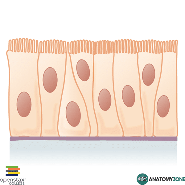
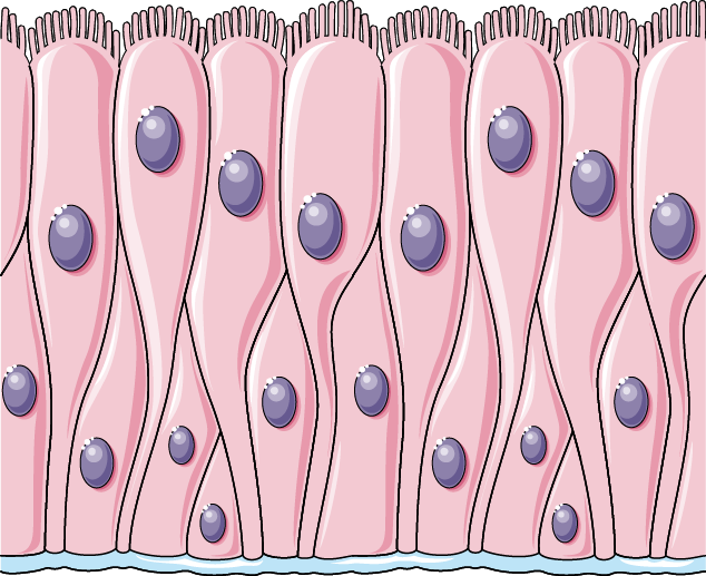
Pseudostratified Ciliated Columnar Epithelium Drawing - Though it is a single layer of cells along the basement membrane, the alignment of the nuclei is not in the same plane and appears as multiple layers. The nucleus is elongated and located on the basal side of the cell. The nuclei of these epithelial cells are at different levels leading to the illusion of being stratified. Drawn by using h & e pencils. Drawing histological diagram of respiratory epithelia.
Smooth (nonciliated tissues) are in the digestive tract, bladder: Though it is a single layer of cells along the basement membrane, the alignment of the nuclei is not in the same plane and appears as multiple layers. Web illustration depicting pseudostratified ciliated columnar epithelium. This epithelium is histologically a simple epithelium even though in a crosssection, it might appear as a. Web drawing pseudostratified ciliated epithelium with goblet cells. This is the 4th video of our series in which we have described. Ciliated tissue lines the trachea and much of the upper respiratory tract:

Pseudostratified Columnar Epithelium Illustration HighRes Vector
Ciliated tissue lines the trachea and much of the upper respiratory tract: Pseudostratified columnar (respiratory epithelium) more fully: Web the epithelium classifies as pseudostratified; Web illustration depicting pseudostratified ciliated columnar epithelium. Smooth (nonciliated tissues) are.
PPT Tissues PowerPoint Presentation, free download ID356154
The stratified epithelium is formed of columnar cell. Smooth (nonciliated tissues) are in the digestive tract, bladder: Drawing histological diagram of respiratory epithelia. All the cells are attached to the underlying basement membrane, but the.
Pseudostratified Columnar Epithelium • General • AnatomyZone
Web human respiratory tract the bar in this image shows the thickness of the layer of ciliated pseudostratified epithelium. Sweat glands, salivary glands, and mammary. All the cells are attached to the underlying basement membrane,.

How to draw Pseudostratified columnar epithelium most easy way
Web the conchae, meatuses, and paranasal sinuses are lined by respiratory epithelium composed of pseudostratified ciliated columnar epithelium (figure 22.5). Web about press copyright contact us creators advertise developers terms privacy policy & safety how.
Pseudostratified columnar epithelium Servier Medical Art
Drawn by using h & e pencils. Web the conchae, meatuses, and paranasal sinuses are lined by respiratory epithelium composed of pseudostratified ciliated columnar epithelium (figure 22.5). Simple columnar or pseudostratified columnar tissue / organ:.

Pseudostratified Ciliated Columnar / / The cells that comprise the
Web about press copyright contact us creators press copyright contact us creators Web human respiratory tract the bar in this image shows the thickness of the layer of ciliated pseudostratified epithelium. Web the pseudostratified columnar.

Illustration depicting Pseudostratified Ciliated Columnar Epithelium
Useful for all medical students. The rest of the tissue below the epithelium is mostly connective tissue, but there are some mucous glands (glandular epithelium) just. Web here, in the pseudostratified columnar hand drawing image,.

Pseudostratified Ciliated Columnar Epithelium Canvas Print / Canvas Art
Web pseudostratified ciliated columnar epithelium containing goblet cells lines most of the major airways. This is an example of pseudostratified epithelium, from the trachea. Web the epithelium classifies as pseudostratified; Though it is a single.

Pseudostratified, Ciliated, Columnar Epithelium Slides VWR
Though it is a single layer of cells along the basement membrane, the alignment of the nuclei is not in the same plane and appears as multiple layers. Web the pseudostratified columnar epithelium is a.
Pseudostratified columnar ciliated epithelium with goblet cells and
Sweat glands, salivary glands, and mammary. All the columnar cells within the tissue remain in contact with the basement membrane and thus pseudostratified ciliated columnar epithelium is actually a simple tissue. Web columnar epithelia, which.
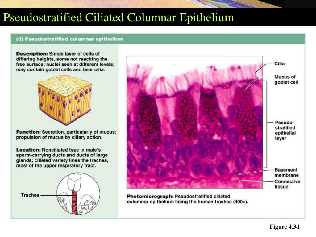
Pseudostratified Ciliated Columnar Epithelium Drawing The trachea is lined with pseudostratified ciliated columnar epithelium cells with goblet cells that produce mucus. Both simple and pseudostratified columnar epithelia are heterogeneous epithelia because they include additional types of cells interspersed among the epithelial cells. Web here, in the pseudostratified columnar hand drawing image, all structures like cells, nucleus, goblet cells, basal cells, and cilia are presented clearly. Web simple columnar epithelium. Pseudostratified ciliated columnar epithelium with goblet cells tissue / organ: