Simple Squamous Epithelium Under Microscope Drawing
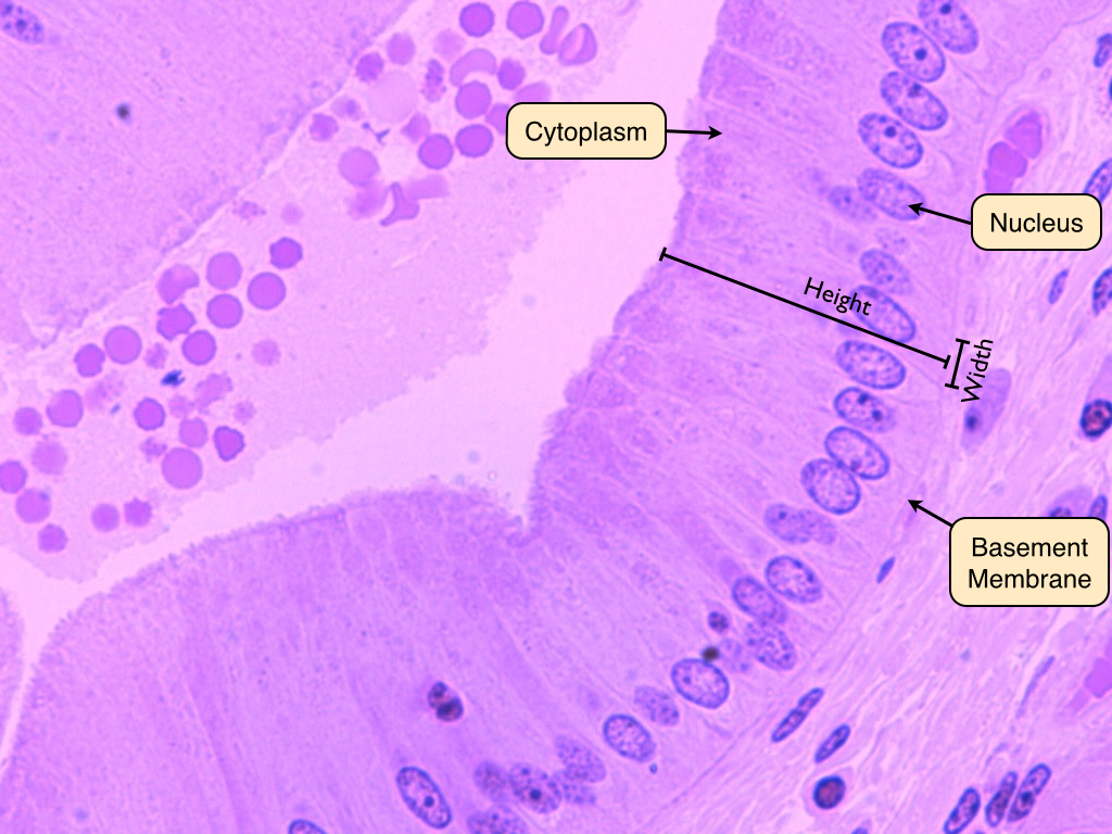
Simple Squamous Epithelium Under Microscope Drawing - The shape of the cells in the single cell layer of simple epithelium reflects the functioning of those cells. Like other epithelial cells, they have polarity and contain a distinct apical surface with specialized membrane proteins. A few epithelial layers are constructed from cells that are said to have a transitional shape. The basement membrane is a thin but strong, acellular layer which lies between the epithelium and the adjacent connective tissue. Now, let’s see the features of stratified squamous epithelium under a microscope with a labeled diagram.
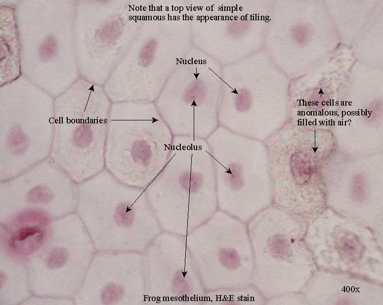
Both the endothelial lining of blood vessels and the mesothelial lining of the body cavities are simple squamous epithelium. Simple squamous epithelia consist of a single layer of flattened cells. Depending on its location, this type of epithelium can function to line and protect an organ or participate in absorption and secretion. A columnar epithelial cell looks like a column or a tall rectangle. Web a squamous epithelial cell looks flat under a microscope. Here, i will provide both hand drawings and real microscope figures of stratified squamous epithelium (keratinized and nonkeratinized). This type of epithelia lines the inner surface of all blood vessels (endothelium), forms the wall of alveolar sacs in the lung.

Human Simple Squamous Epithelium, sec. 7 µm, H&E Microscope Slide
Squamous cell nuclei tend to be flat, horizontal, and elliptical, mirroring the form of the cell. Web simple squamous epithelium, because of the thinness of the cell, is present where rapid passage of chemical compounds.

Simple Squamous Epithelial Tissue Under Microscope
Web simple squamous epithelium, because of the thinness of the cells, is present where rapid passage of chemical compounds is necessary such as the lining of capillaries and the small air sacs of the lung..
Simple Squamous Epithelium Inrtroducrion , Anatomy & Function
Try to identify the simple squamous epithelia in these pictures. Web simple squamous epithelia are tissues formed from one layer of squamous cells that line surfaces. The nucleus looks like the yolk. Web a simple.

Labeled Simple Squamous Epithelium Under Microscope 400x Micropedia
Web simple squamous epithelium, because of the thinness of the cells, is present where rapid passage of chemical compounds is necessary such as the lining of capillaries and the small air sacs of the lung..

simple squamous epithelium Histología, Biología, Anatomia patologica
Web the simple squamous epithelium location specifically exists in the lining of the blood vessels like the arteries, veins, and capillaries. Here, i will provide both hand drawings and real microscope figures of stratified squamous.

Simple Squamous Epithelium Location And Function Steve Gallik
Web there are three basic shapes used to classify epithelial cells. Now, let’s see the features of stratified squamous epithelium under a microscope with a labeled diagram. The dark purple spots are the nuclei of.

Simple Squamous Epithelium 40X Annotated Histology
Web introduction the first pages illustrate introductory concepts for those new to microscopy as well as definitions of commonly used histology terms. The basement membrane is a thin but strong, acellular layer which lies between.
Epithelial Tissue Anatomy & Physiology
Web simple squamous epithelium is a type of simple epithelium that is formed by a single layer of cells on a basement membrane. The cells in simple squamous epithelium have the appearance of thin scales..

Simple Squamous Epithelium Diagram Quizlet
The nucleus looks like the yolk. A cuboidal epithelial cell looks close to a square. Simple squamous epithelia consist of a single layer of flattened cells. It is also found lining the alveoli or air.

How to draw stratified squamous epithelium easy way YouTube
Web simple squamous epithelium identification points function and location of simple epithelium simple cuboidal epithelium histology functions and location of simple cuboidal epithelial tissue identification of simple columnar epithelium functions of simple columnar epithelium and.
Simple Squamous Epithelium Under Microscope Drawing A few epithelial layers are constructed from cells that are said to have a transitional shape. Web simple squamous epithelium identification points function and location of simple epithelium simple cuboidal epithelium histology functions and location of simple cuboidal epithelial tissue identification of simple columnar epithelium functions of simple columnar epithelium and their location identification of stratified squamous epithelium It is also found lining the alveoli or air sacs within the. The alveoli of lungs where gases diffuse, segments of kidney tubules, and the lining of capillaries are also made of simple squamous epithelial tissue. The cells in simple squamous epithelium have the appearance of thin scales.