Spinal Cord Drawing
Spinal Cord Drawing - Vector sketch icons of human body bones and joints. Web according to its rostrocaudal location the spinal cord can be divided into four parts: Web use this line drawing to refresh your understanding of the gross anatomy of the spinal cord, paying particular attention to the following structures in this figure: Diagram of the spinal cord is illustrated in detail with neat and clear labelling. Human joints and body parts bones sketch icons.
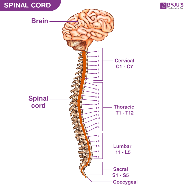
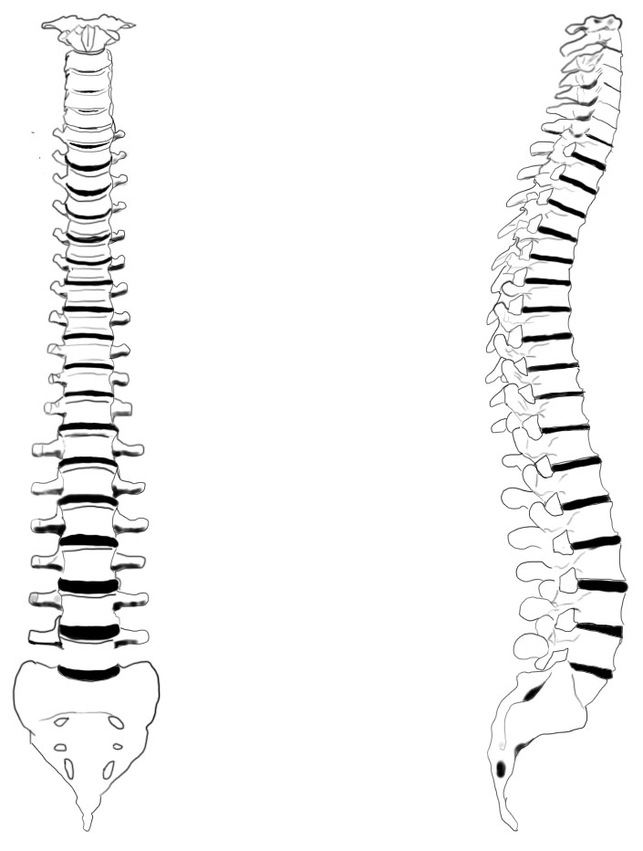
Spinal cord drawing stock photos are available in a variety of sizes and formats to fit your needs. Cervical enlargement, lumbosacral enlargement, medullary cone, spinal part of filum terminale, cauda equina, spinal nerves. Here i upload carefully crafted videos to meet the problems in drawing.all the videos are. Let us learn how to draw a human. Web here's a drawing of the spinal cord. Cervical, thoracic, lumbar and sacral, two of these are marked by an upper (cervical) and a lower (lumbar) enlargement. Learn more about the spinal cord with our learning material.

11.1A Overview of the Spinal Cord Medicine LibreTexts
Web the module promotes learning and mastery of spinal cord anatomy and lesion localization. And there's a number of structures coming out of the spinal cord that i'll talk about next. Web according to its.

Spinal Cord Anatomy Nurse Info
A number of approaches exist to improve learning and retention of neuroanatomy and clinical localization principles. Web here's a drawing of the spinal cord. These nerve signals help you feel sensations and move your muscles..
Spinal cord diagram
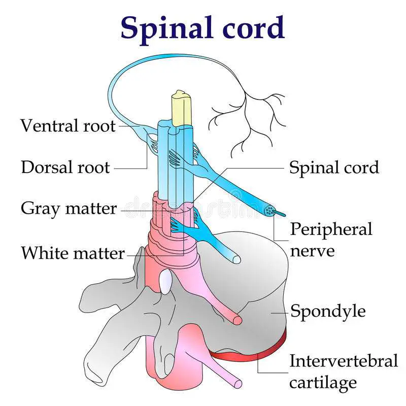
From the spinal cord se dã©tachent on each side rachidian nerves constituted of a ganglion, a posterior and an anterior root. Representation in 3/4 front view of the stucture of the spinal cord, and rachidian.

Anatomy and Health Charts Free Printable PDF Files Human body anatomy
This kind of long tube that runs down the spine. Ventral horn, dorsal horn, white matter, gray matter, meninges, central canal, dorsal root ganglion, dorsal root of the spinal nerve, and the ventral root of.

How Does The Spinal Cord Work Reeve Foundation
Web the module promotes learning and mastery of spinal cord anatomy and lesion localization. Welcome to my just made easy official youtube channel. Web the module promotes learning and mastery of spinal cord anatomy and.
Spinal Cord Anatomy, Structure, Function, & Diagram
Web your spinal cord is the long, cylindrical structure that connects your brain and lower back. Web use this line drawing to refresh your understanding of the gross anatomy of the spinal cord, paying particular.
The spinal cord Anatomy of the spinal cord Physiology of the spinal
Web vector human spine blueprint. Vector sketch icons of human body bones and joints. Ventral horn, dorsal horn, white matter, gray matter, meninges, central canal, dorsal root ganglion, dorsal root of the spinal nerve, and.
Anatomy of the Spinal Cord Praxis Spinal Cord Institute
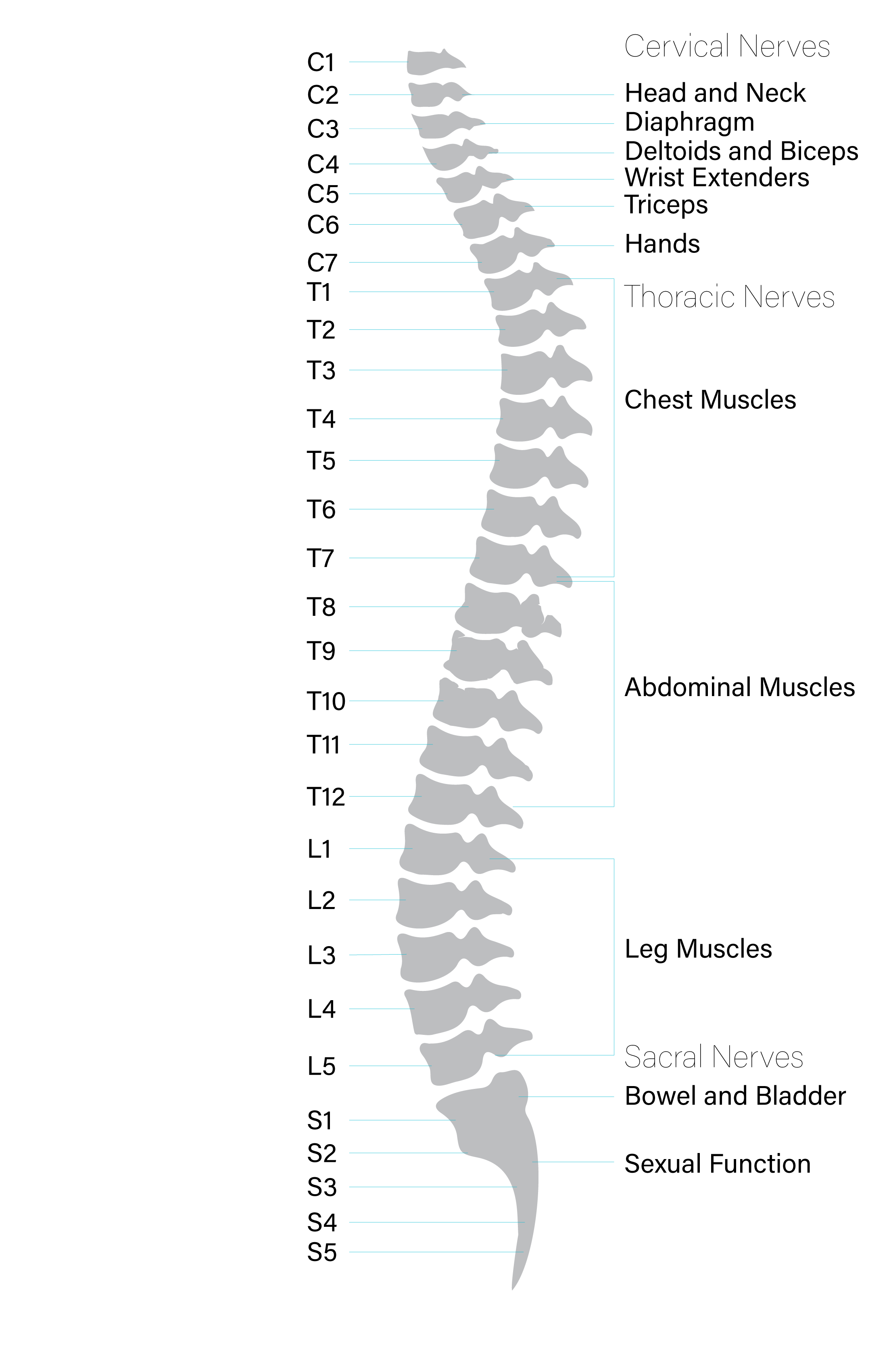
Many of the nerves of the peripheral nervous system, or pns, branch out from the spinal cord and travel to. Diagram of the spinal cord is illustrated in detail with neat and clear labelling. Web.
Human Spinal Cord Drawing Sketch Coloring Page
Spine isolated on a white backgrounds. And there's a number of structures coming out of the spinal cord that i'll talk about next. These nerve signals help you feel sensations and move your muscles. Web.

The Spinal Cord Neurologic Clinics
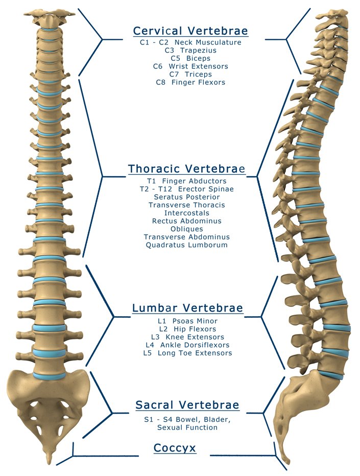
A bony column of vertebrae surrounds and protects your spinal cord. Web the module promotes learning and mastery of spinal cord anatomy and lesion localization. Here i upload carefully crafted videos to meet the problems.
Spinal Cord Drawing Ventral horn, dorsal horn, white matter, gray matter, meninges, central canal, dorsal root ganglion, dorsal root of the spinal nerve, and the ventral root of the spinal nerve. Web each segment of the spinal cord provides several pairs of spinal nerves, which exit from vertebral canal through the intervertebral foramina. Web the module promotes learning and mastery of spinal cord anatomy and lesion localization. Web the module promotes learning and mastery of spinal cord anatomy and lesion localization. Web according to its rostrocaudal location the spinal cord can be divided into four parts: