Whitefish Blastula Mitosis Drawing
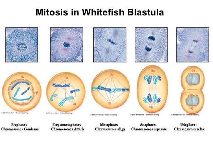
Whitefish Blastula Mitosis Drawing - View the slide on the objective which provides the best view. Obtain a slide of a whitefish blastula. Whitefish blastula a blastula is an early stage of embryonic development of animals. Web since early embryogenesis involves rapid cellular division, the whitefish blastula has long served as a model of mitotic division in animals. Interphase, prophase, metaphase, anaphase, and telophase.
Web the cells of a developing embryo are dividing rapidly and can be used for viewing the different stages of mitosis. Web observe the prepared slide of a whitefish blastula under high power (400x). Onion root tip whitefish blastula; Use the image slider below to learn how to use a microscope to identify cells dividing by mitosis on a whitefish embryo slide. Then draw cells in cytokinesis and interphase as well. Events & activities of each + slide of whitefish blastula slides learn with flashcards, games, and more — for free. Mitosis is part of a.

Whitefish Blastula Cells, Mitosis, Lm Photograph by Michael Abbey Pixels
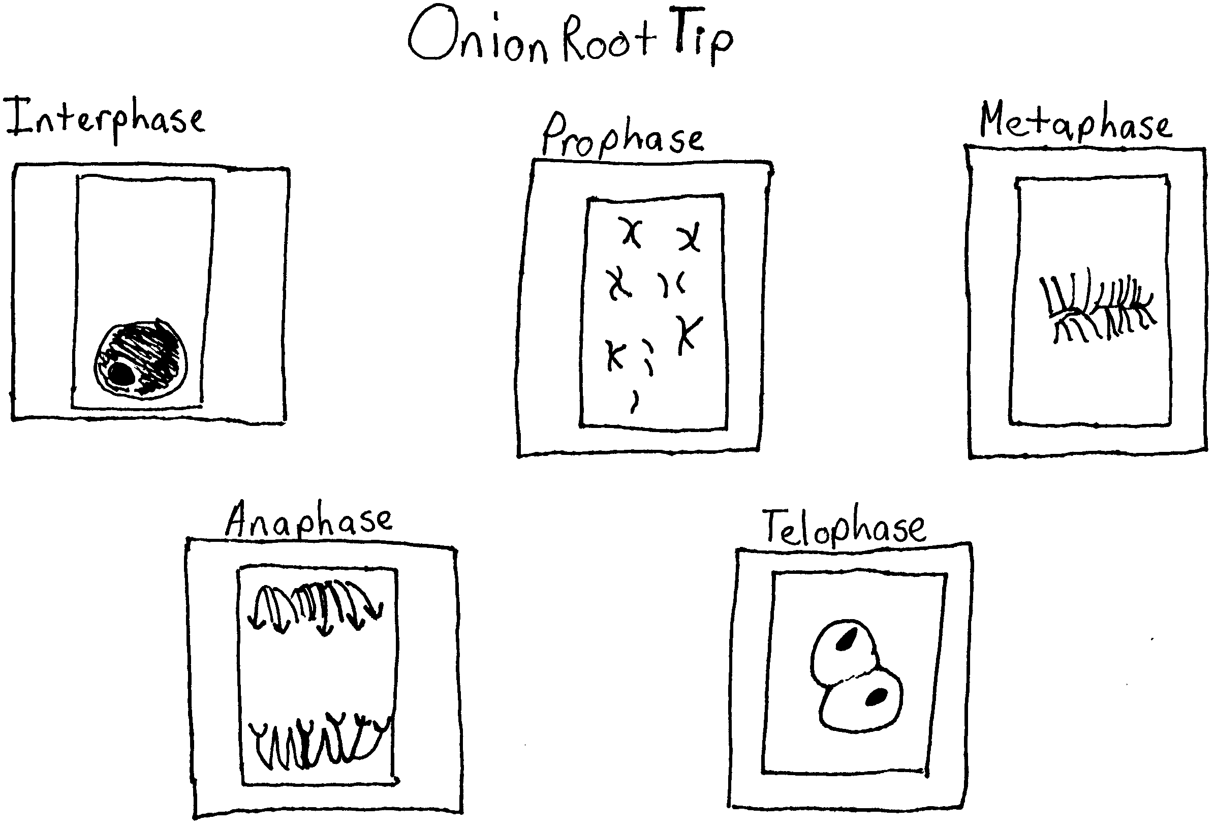
Then draw cells in cytokinesis and interphase as well. Find the six stages of the cell cycle above, and draw and label what you see in each stage on the paper provided. Fall 2005 onion.
Chapter 8 handout blks_ 10182011
Web since early embryogenesis involves rapid cellular division, the whitefish blastula has long served as a model of mitotic division in animals. Web why are whitefish blastula used to study mitosis? Interphase, prophase, metaphase, anaphase,.
Stages of Mitosis in the Blastula of a Whitefish Lab Manual for
It also has the advantage of demonstrating clear spindle formation in the cytoplasm. At this stage, the embryo Observe the stages of mitosis in the blastula of a whitefish a fundamental property of somatic (nonreproductive.
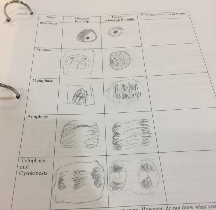
Solved Important Features of Stage Stage Diagram Diagramm
Mitosis is considered nuclear division, since its main stages deal strictly with the nucleus and its contents (dna). Find the six stages of the cell cycle above, and draw and label what you see in.

AP Lab 3 Sample 3 Mitosis BIOLOGY JUNCTION
The blastula is an early stage of embryo development and represents a period in the organism's life when most of the cells are constantly dividing. Web in this chapter, you can use pictures of whitefish.

Whitefish Mitosis Whitefish Embryo And Chromosomes Are Still Visible
The blastula of a whitefish and the root tip of an onion. The blastula is an early stage of embryo development and represents a period in the organism's life when most of the cells are.

Whitefish Mitosis Telophase Cytokinesis Embryo Still Visible 400x 56
Draw a table in which you contrast mitosis in onions and whitefish. Then draw cells in cytokinesis and interphase as well. Obtain a slide of a whitefish blastula. Whitefish blastula a blastula is an early.
😎 Whitefish interphase. Mitosis Whitefish Blastula Flashcards. 20190201
Draw a cell in anaphase. The student will correctly identify and draw four stages of mitosis using microscope slide images of onion root tips and whitefish blastulae. Then draw cells in cytokinesis and interphase as.

Mitotic cell division stages of Whitefish blastula YouTube
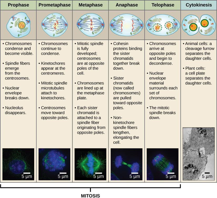
Web cytokinesis begins at anaphase and continues through and beyond telophase. Mitosis consists of 4 major stages: Interphase, prophase, metaphase, anaphase, and telophase. Nuclear membrane breaks down, chromatin condenses, mitotic spindle forms and attaches to.

Whitefish mitosis Diagram Quizlet
Web obtain a slide of a whitefish embryo (blastula) from the slide box at your table. Use the image slider below to learn how to use a microscope to identify cells dividing by mitosis on.
Whitefish Blastula Mitosis Drawing Find the six stages of the cell cycle above, and draw and label what you see in each stage on the paper provided. Examine the slide under a microscope. Prophase, metaphase, anaphase, and telophase. Web two specimens are commonly used by biologists to study mitosis: The whitefish embryo is a good place to look at mitosis because these cells are rapidly dividing as the fish embryo is growing.