Skull X Ray

The use of X-ray technology in medical imaging has revolutionized the field of diagnostics, allowing healthcare professionals to visualize internal structures of the body in detail. One of the most critical applications of X-ray technology is in the imaging of the skull, which can help diagnose a range of conditions, from fractures and tumors to vascular abnormalities and congenital disorders.
Introduction to Skull X-Ray
A skull X-ray is a non-invasive medical imaging procedure that uses X-rays to produce detailed images of the skull, including the bones, blood vessels, and soft tissues. The procedure is typically performed in a hospital or radiology department, and the resulting images are interpreted by a radiologist or other medical specialist. Skull X-rays can be used to diagnose a range of conditions, including:
- Fractures: Skull X-rays can help diagnose fractures, including linear, depressed, and basilar skull fractures.
- Tumors: Skull X-rays can help diagnose tumors, including benign and malignant types, such as meningiomas and osteomas.
- Vascular abnormalities: Skull X-rays can help diagnose vascular abnormalities, including aneurysms and arteriovenous malformations.
- Congenital disorders: Skull X-rays can help diagnose congenital disorders, including craniosynostosis and plagiocephaly.
How Skull X-Ray Works
The process of undergoing a skull X-ray is relatively straightforward. The patient is typically positioned on an X-ray table, and the X-ray machine is adjusted to capture images of the skull from various angles. The X-ray machine emits a low-level radiation beam that passes through the skull, and the resulting images are captured on a digital detector or film. The entire procedure typically takes several minutes to complete, and the patient may be required to hold still or change positions to capture images from different angles.
Types of Skull X-Ray
There are several types of skull X-ray procedures, including:
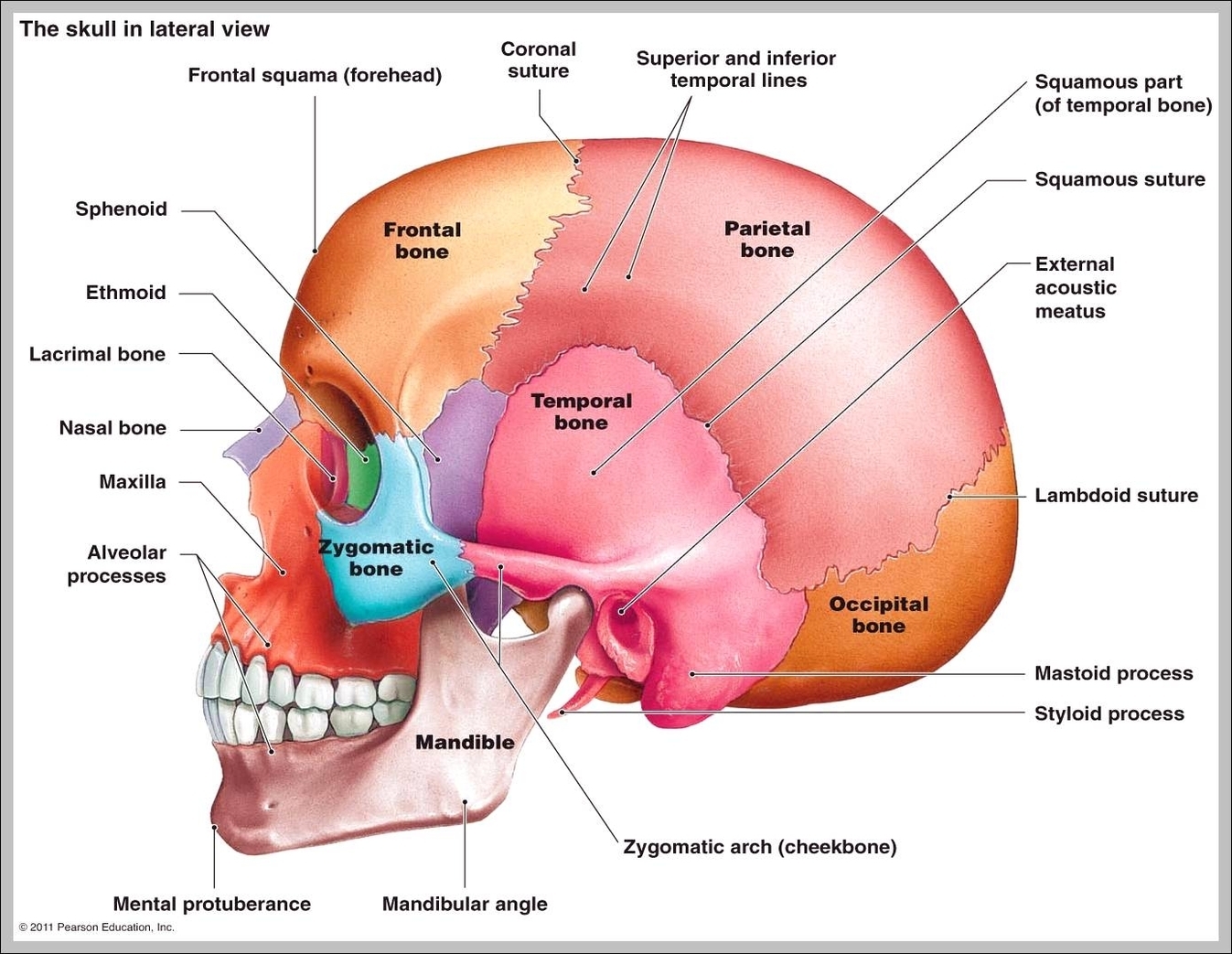
- Lateral skull X-ray: This type of X-ray captures images of the skull from the side, and is often used to diagnose fractures and tumors.
- Anteroposterior (AP) skull X-ray: This type of X-ray captures images of the skull from the front to the back, and is often used to diagnose vascular abnormalities and congenital disorders.
- Towne’s view: This type of X-ray captures images of the skull from an angle, and is often used to diagnose fractures and tumors.
Interpreting Skull X-Ray Results
The results of a skull X-ray are typically interpreted by a radiologist or other medical specialist, who will examine the images for signs of abnormality. The interpreter will look for signs of fractures, tumors, vascular abnormalities, and congenital disorders, and will also assess the overall health of the skull and surrounding tissues. In some cases, additional imaging tests, such as CT or MRI scans, may be required to confirm a diagnosis or gather more detailed information.
It's essential to note that skull X-rays are just one tool in the diagnostic toolkit, and should be used in conjunction with other imaging modalities and clinical evaluations to confirm a diagnosis. A radiologist or other medical specialist should always interpret the results of a skull X-ray, as they have the training and expertise to accurately identify signs of abnormality.
Advantages and Disadvantages of Skull X-Ray
Skull X-rays have several advantages, including:
- Non-invasive: Skull X-rays are a non-invasive procedure, which means that they do not require surgery or the insertion of instruments into the body.
- Quick and easy: Skull X-rays are a relatively quick and easy procedure, which can be completed in several minutes.
- Low cost: Skull X-rays are a relatively low-cost procedure, compared to other imaging modalities such as CT or MRI scans.
However, skull X-rays also have some disadvantages, including:
- Limited detail: Skull X-rays may not provide as much detail as other imaging modalities, such as CT or MRI scans.
- Radiation exposure: Skull X-rays involve exposure to low-level radiation, which may be a concern for some patients.
- Limited sensitivity: Skull X-rays may not be sensitive enough to detect certain types of abnormalities, such as small fractures or tumors.
Real-World Applications of Skull X-Ray
Skull X-rays have a range of real-world applications, including:
- Emergency medicine: Skull X-rays are often used in emergency medicine to quickly diagnose fractures and other injuries.
- Neurosurgery: Skull X-rays are often used in neurosurgery to plan surgical procedures and diagnose tumors and vascular abnormalities.
- Pediatrics: Skull X-rays are often used in pediatrics to diagnose congenital disorders and other conditions that affect the developing skull.
Skull X-rays are a valuable diagnostic tool that can help healthcare professionals diagnose a range of conditions, from fractures and tumors to vascular abnormalities and congenital disorders. While they have some limitations, skull X-rays are a non-invasive, quick, and easy procedure that can provide valuable information about the health of the skull and surrounding tissues.
Frequently Asked Questions
What is a skull X-ray used for?
+A skull X-ray is used to diagnose a range of conditions, including fractures, tumors, vascular abnormalities, and congenital disorders.
How long does a skull X-ray take?
+A skull X-ray typically takes several minutes to complete.
Is a skull X-ray safe?
+Skull X-rays involve exposure to low-level radiation, but the benefits of the procedure typically outweigh the risks.
In conclusion, skull X-rays are a valuable diagnostic tool that can help healthcare professionals diagnose a range of conditions. While they have some limitations, skull X-rays are a non-invasive, quick, and easy procedure that can provide valuable information about the health of the skull and surrounding tissues. By understanding the advantages and disadvantages of skull X-rays, healthcare professionals can use this technology to improve patient outcomes and advance our understanding of the human body.


