X Ray Of Abdomen: Get Accurate Injury Detection

The abdominal region, often referred to as the abdomen, is a complex and vital part of the human body. It houses several crucial organs, including the liver, stomach, small intestine, kidneys, and pancreas, among others. Due to its complex nature and the importance of its contents, diagnosing injuries or conditions in this area can be challenging. One of the primary diagnostic tools used for examining the abdomen is the X-ray. An X-ray of the abdomen is a non-invasive medical imaging technique used to produce images of the internal structures of the abdominal region.
How X-Ray Works for Abdominal Injury Detection
X-rays are a form of electromagnetic radiation that can penetrate solid objects, including the human body. When an X-ray is taken, the machine sends X-ray beams through the body, and these beams are absorbed or deflected by various tissues and structures within. Denser materials like bones absorb more X-rays, appearing white on the image, while softer tissues absorb fewer, appearing in shades of gray. Air-filled spaces, like the lungs or the gastrointestinal tract if it contains gas, appear black because X-rays pass through them with minimal absorption.
For detecting abdominal injuries, an X-ray can be particularly useful for identifying certain types of issues, such as:
- Foreign objects: If a patient has ingested or otherwise introduced a foreign object into their body, an X-ray can often detect it, especially if the object is metallic or dense.
- Fractures: While not the primary method for diagnosing abdominal organ injuries, X-rays can reveal fractures of the ribs, spine, or pelvis that might be associated with abdominal trauma.
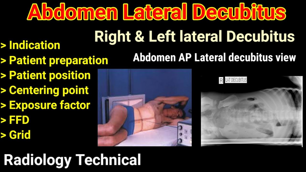
- Free air: In cases of perforation of the gastrointestinal tract, free air can be detected under the diaphragm, indicating a serious condition that requires immediate medical attention.
- Obstructions or blockages: Sometimes, an X-ray can suggest the presence of an obstruction in the intestine by showing dilated loops of bowel.
Preparation and Procedure
Preparing for an abdominal X-ray typically involves removing any clothing or jewelry that might interfere with the X-ray beams. Depending on the reason for the X-ray, patients might be asked to change into a hospital gown. The procedure itself is relatively quick and straightforward:
- Positioning: The patient is positioned on an X-ray table, either standing, sitting, or lying down, depending on the type of X-ray being taken and the patient’s condition.
- X-ray machine adjustment: The X-ray machine is adjusted according to the patient’s size and the area to be imaged.
- Exposure: The X-ray beam is directed at the abdominal area for a few seconds. The patient will be asked to hold their breath and remain still to ensure a clear image.
- Image review: The radiologist reviews the images for any signs of injury or abnormality.
Limitations and Alternatives
While X-rays are valuable for detecting certain types of abdominal injuries, they have limitations. For example, they are not effective at visualizing soft tissue injuries, such as damage to organs like the liver, spleen, or kidneys. In such cases, other imaging techniques might be used, including:
- Computed Tomography (CT) scans: Provide detailed images of both bone and soft tissue.
- Magnetic Resonance Imaging (MRI): Excellent for visualizing soft tissues, though not always the first choice in emergency situations due to longer scanning times and higher costs.
- Ultrasound: Can be useful for evaluating certain abdominal injuries, especially in emergency settings or when CT scans are not readily available.
Conclusion
An X-ray of the abdomen is a fundamental tool in the diagnostic arsenal for detecting certain types of injuries or conditions in the abdominal region. While it has its limitations, especially concerning soft tissue visualization, it remains a crucial first step in many diagnostic pathways due to its speed, availability, and non-invasive nature. For comprehensive evaluation, especially in complex trauma cases, it is often used in conjunction with other diagnostic imaging modalities.
What is the primary use of an abdominal X-ray in injury detection?
+The primary use of an abdominal X-ray is to quickly identify certain types of injuries or conditions within the abdominal cavity, such as the presence of foreign objects, free air under the diaphragm indicating a perforation, or obstructions in the intestine.
Are there any alternatives to X-rays for detecting abdominal injuries?
+Yes, alternatives include CT scans for detailed imaging of both bone and soft tissue, MRI for excellent soft tissue visualization, and ultrasound for evaluating certain conditions, especially in emergency settings or when other methods are not available.
How does an X-ray detect issues in the abdominal region?
+An X-ray detects issues by using electromagnetic radiation that penetrates the body. Different tissues absorb X-rays at varying levels, allowing for the visualization of internal structures. Denser materials like bones appear white, while softer tissues appear in shades of gray, and air-filled spaces appear black.
In summary, while an X-ray of the abdomen provides valuable information for certain types of injuries, a comprehensive diagnostic approach often involves combining X-ray findings with those from other imaging modalities to ensure accurate detection and appropriate treatment of abdominal injuries.


