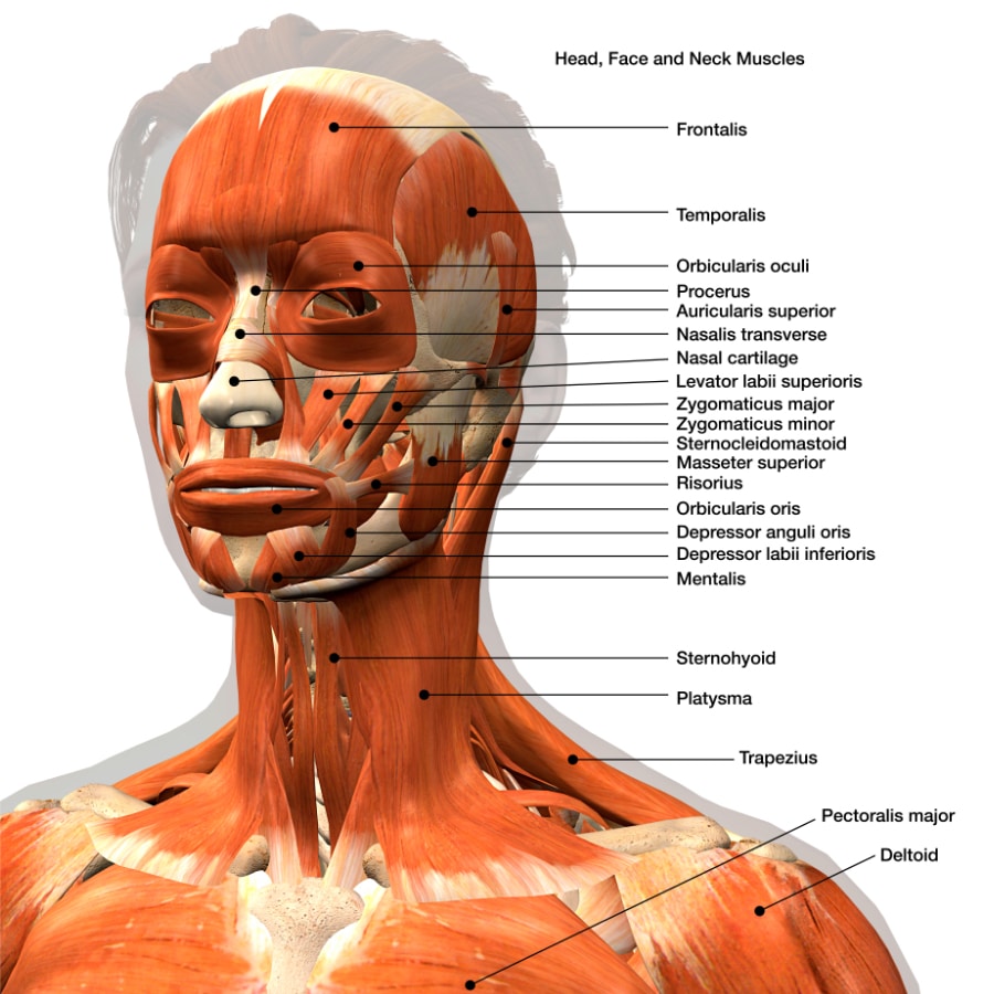
Head Mri Pictures

Magnetic Resonance Imaging (MRI) of the head is a non-invasive diagnostic technique that provides detailed images of the brain and its surrounding structures. The process involves the use of a strong magnetic field, radio waves, and a computer to generate high-resolution images of the internal structures of the head. These images can be used to diagnose and monitor a wide range of conditions, including tumors, strokes, and neurological disorders.
How Head MRI Works
When a person undergoes a head MRI, they are placed inside a large, cylindrical machine that contains a strong magnet. The magnet produces a magnetic field that aligns the hydrogen atoms in the body. Radio waves are then used to disturb the alignment of these atoms, causing them to emit signals that are picked up by the MRI machine. These signals are used to create detailed images of the internal structures of the head.
Types of Head MRI
There are several types of head MRI that can be used to diagnose and monitor different conditions. Some of the most common types of head MRI include:
- T1-Weighted MRI: This type of MRI is used to produce detailed images of the brain and its surrounding structures. It is particularly useful for diagnosing conditions such as tumors and stroke.
- T2-Weighted MRI: This type of MRI is used to produce images of the brain that highlight areas of inflammation or damage. It is particularly useful for diagnosing conditions such as multiple sclerosis and brain injuries.
- Functional MRI (fMRI): This type of MRI is used to produce images of the brain that show areas of activity. It is particularly useful for diagnosing conditions such as epilepsy and stroke.
- Magnetic Resonance Angiography (MRA): This type of MRI is used to produce images of the blood vessels in the head. It is particularly useful for diagnosing conditions such as aneurysms and vascular malformations.
What to Expect During a Head MRI
When a person undergoes a head MRI, they can expect the following:
- Preparation: The person will be asked to remove any metal objects, such as jewelry or glasses, and change into a hospital gown.
- Positioning: The person will be placed on a table that slides into the MRI machine.
- Scanning: The MRI machine will produce a loud knocking or banging sound as it takes images of the head.
- Contrast: A contrast agent may be injected into the person’s vein to highlight certain areas of the head.
- Length of procedure: The procedure can take anywhere from 15 to 90 minutes, depending on the type of MRI and the number of images needed.
Interpreting Head MRI Pictures
Head MRI pictures are interpreted by a radiologist, who looks for any abnormalities or areas of concern. The radiologist will examine the images for signs of conditions such as tumors, stroke, and neurological disorders. The radiologist will also look for any signs of damage or inflammation in the brain and its surrounding structures.
It's essential to note that head MRI pictures should only be interpreted by a qualified radiologist. The images can be complex and require specialized training to interpret accurately.
Common Conditions Diagnosed with Head MRI
Head MRI is used to diagnose and monitor a wide range of conditions, including:
- Tumors: Head MRI can be used to diagnose and monitor brain tumors, such as glioblastoma and meningioma.
- Stroke: Head MRI can be used to diagnose and monitor stroke, including ischemic and hemorrhagic stroke.
- Neurological disorders: Head MRI can be used to diagnose and monitor neurological disorders, such as multiple sclerosis, Parkinson’s disease, and Alzheimer’s disease.
- Brain injuries: Head MRI can be used to diagnose and monitor brain injuries, such as concussions and traumatic brain injuries.
Pros and Cons of Head MRI
- Pros:
- Non-invasive and painless
- Provides detailed images of the brain and its surrounding structures
- Can be used to diagnose and monitor a wide range of conditions
- Cons:
- Can be expensive
- May require contrast agent
- Can be claustrophobic for some people
Conclusion
Head MRI is a powerful diagnostic tool that provides detailed images of the brain and its surrounding structures. It is used to diagnose and monitor a wide range of conditions, including tumors, stroke, and neurological disorders. By understanding how head MRI works and what to expect during the procedure, individuals can feel more comfortable and confident in the diagnostic process.
What is the difference between a head MRI and a head CT scan?
+A head MRI uses a strong magnetic field and radio waves to produce detailed images of the brain and its surrounding structures, while a head CT scan uses X-rays to produce images of the brain and its surrounding structures.
How long does a head MRI take?
+The length of a head MRI can vary depending on the type of MRI and the number of images needed. On average, a head MRI can take anywhere from 15 to 90 minutes.
Is a head MRI painful?
+No, a head MRI is not painful. However, some people may experience claustrophobia or discomfort during the procedure.


